Anat Cell Biol.
2016 Dec;49(4):249-253. 10.5115/acb.2016.49.4.249.
An osteological study of supratrochlear foramen of humerus of south Indian population with reference to anatomical and clinical implications
- Affiliations
-
- 1Department of Anatomy, Sri Siddhartha Medical College, Tumkur, India. drshivaleela83@yahoo.co.in
- KMID: 2365554
- DOI: http://doi.org/10.5115/acb.2016.49.4.249
Abstract
- The supratrochlear foramen (STF) is an important and relatively common anatomic variation in the lower end of the humerus in humans. The present study on south Indian population is an attempt to highlight the incidence, morphological features and clinical importance of STF. The study was conducted on dried human humeri of unknown sex and free of pathological changes. The presence of a STF was noted for its shape and divided into three types (oval, round, and irregular). In bones where the foramen was absent the translucency of the septum between the coronoid and the radial fossa was noted by placing the lower end of the humerus against the X-ray view box. Out of the 142 humeri studied, 72 humeri (50.7%) were right sided and 70 humeri (49.2%) were left sided. In these 142 humeri, 38 humeri (26.7%) showed the presence of STF. The majority of STF were round shaped in 47.37%, followed by oval shaped in 42.11% and 10.53% were irregular in shape. The STF was absent in 36 humeri (25.35%) and 68 humeri (47.89%) showed the translucency of septum. The mean transverse diameter on right side was 4.50±3.183 mm and 3.32±3.222 mm on left side. The mean vertical diameter was 3.88±2.391 on right side and 3.68±3.532 mm on left side. Its existence is important to the orthopaedician in the preoperative planning of nailing fractures of the distal humerus and to the radiologist for differentiating it from an osteolytic or cystic lesion.
Figure
-
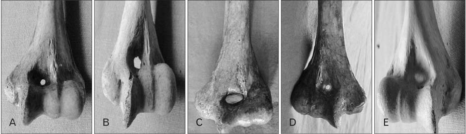
Fig. 1 Photograph showing the various shapes of supratrochlear foramen (STF). (A) Round STF. (B) Irregular STF. (C) Oval STF. (D) STF with translucent septum. (E) Translucent septum.
Reference
-
1. Kate BR, Dubey PN. A note on the septal apertures in the humerus of Central Indians. East Anthropol. 1970; 33:105–110.2. Akabori E. Septal apertures in the humerus in Japanese, Ainu and Koreans. Am J Phys Anthropol. 1934; 18:395–400.3. Das S. Supratrochlear foramen of the humerus. Anat Sci Int. 2008; 83:120.4. Morton HS, Crysler WE. Osteochondritis dissecans of the supratrochlear septum. J Bone Joint Surg Am. 1945; 27:12–24.5. De Wilde V, De Maeseneer M, Lenchik L, Van Roy P, Beeckman P, Osteaux M. Normal osseous variants presenting as cystic or lucent areas on radiography and CT imaging: a pictorial overview. Eur J Radiol. 2004; 51:77–84.6. Haziroglu RM, Ozer M. A supratrochlear foramen in the humerus of cattle. Anat Histol Embryol. 1990; 19:106–108.7. Benfer RA, Tappen NC. The occurrence of the septal perforation of the humerus in three non-human primate species. Am J Phys Anthropol. 1968; 29:19–28.8. Singhal S, Rao V. Supratrochlear foramen of the humerus. Anat Sci Int. 2007; 82:105–107.9. Chapman DL, Garvey N, Hancock S, Alexiou M, Agulnik SI, Gibson-Brown JJ, Cebra-Thomas J, Bollag RJ, Silver LM, Papaioannou VE. Expression of the T-box family genes, Tbx1-Tbx5, during early mouse development. Dev Dyn. 1996; 206:379–390.10. Govoni KE, Linares GR, Chen ST, Pourteymoor S, Mohan S. T-box 3 negatively regulates osteoblast differentiation by inhibiting expression of osterix and runx2. J Cell Biochem. 2009; 106:482–490.11. Paraskevas GK, Papaziogas B, Tzaveas A, Giaglis G, Kitsoulis P, Natsis K. The supratrochlear foramen of the humerus and its relation to the medullary canal: a potential surgical application. Med Sci Monit. 2010; 16:BR119–BR123.12. Nayak SR, Das S, Krishnamurthy A, Prabhu LV, Potu BK. Supratrochlear foramen of the humerus: an anatomico-radiological study with clinical implications. Ups J Med Sci. 2009; 114:90–94.13. Ndou R, Smith P, Gemell R, Mohatla O. The supratrochlear foramen of the humerus in a South African dry bone sample. Clin Anat. 2013; 26:870–874.14. Glanville EV. Perforation of the coronoid-olecranon septum. Humero-ulnar relationships in Netherlands and African populations. Am J Phys Anthropol. 1967; 26:85–92.15. Hirsh IS. The supratrochlear foramen: clinical and anthropological considerations. Am J Surg. 1927; 2:500–505.16. Warren E. An investigation on the variability of the human skeleton with especial reference to the naqada race, discovered by professor Flinders Petrie in his explorations in Egypt. Proc R Soc Lond. 1897; 61:398–401.17. Macalister A. Anatomical notes and quaries. Series II. 1. Perforate humeri in ancient Egyptian skeletons. J Anat Phys. 1990; 35:121–122.18. Papaloucas C, Papaloucas M, Stergioulas A. Rare cases of humerus septal apertures in Greeks. Trends Med Res. 2011; 6:178–183.19. Öztürk A, Kutlu C, Bayraktar B, Ari Z, Sahinoglu K. The supratrochlear foramen in the humerus: anatomical study. Istanbul Tip Fak Mecmuasi. 2000; 63:72–76.20. Chatterjee KP. The incidence of perforation of olecranon fossa in the humerus among Indians. East Anthropol. 1968; 21:279–284.21. Singh S, Singh SP. A study of the supratrochlear foramen in the humerus of North Indians. J Anat Soc India. 1972; 21:52–56.22. Çimen M, Koşar Y, Sönmez M. Humerus' ta apertura septalis ile ilgili bir araştırma. Antropoloji. 2003; 14:20–23.23. Ming-Tzu P. Septal apertures in the humerus in the Chinese. Am J Phys Anthropol. 1935; 20:165–170.24. Krishnamurthy A, Yelicharla AR, Takkalapalli A, Munishamappa V, Bovinndala B, Chandramohan M. Supratrochlear foramen of humerus: a morphometric study. Int J Biol Med Res. 2011; 2:829–831.25. Hrdlička A. The humerus: septal apertures. Anthropology. 1932; 10:31–96.26. Benfer RA, McKern TW. The correlation of bone robusticity with the perforation of the coronoid-olecranon septum in the humerus of man. Am J Phys Anthropol. 1966; 24:247–252.27. Veerappan V, Ananthi S, Kannan NG, Prabhu K. Anatomical and radiological study of supratrochlear foramen of humerus. World J Pharm Pharm Sci. 2013; 2:313–320.28. Mathew AJ, Gopidas GS, Sukumaran TT. A study of the supratrochlear foramen of the humerus: anatomical and clinical perspective. J Clin Diagn Res. 2016; 10:AC05–AC08.29. Cheng JC, Shen WY. Limb fracture pattern in different pediatric age groups: a study of 3,350 children. J Orthop Trauma. 1993; 7:15–22.30. Sunday OO, Olusegun OS, Oluwabunmi BM. The supratrochlear foramen of the humerus: implications for intramedullary nailing in distal humerus. J Biol Agric Healthc. 2014; 4:2224–3208.
- Full Text Links
-
- Actions
-
Cited
- CITED
-
- Close
- Share
-
- Similar articles
-
- Morphology and topography of the parietal emissary foramina in South Indians: an anatomical study
- Morphometric study on mandibular foramen and incidence of accessory mandibular foramen in mandibles of south Indian population and its clinical implications in inferior alveolar nerve block
- Morphometric analysis of infraorbital foramen in Indian dry skulls
- Sex determination using humeral dimensions in a sample from KwaZulu-Natal: an osteometric study
- A giant foramen of Vesalius: case report
