Chordoid Glioma with Intraventricular Dissemination: A Case Report with Perfusion MR Imaging Features
- Affiliations
-
- 1Department of Radiology, Chonnam National University Medical School, Chonnam National University Hospital, Gwangju 61469, Korea. radyoon@jnu.ac.kr
- 2Department ofForensic Medicine, Chonnam National University Medical School, Chonnam National University Hospital, Gwangju 61469, Korea.
- KMID: 2351173
- DOI: http://doi.org/10.3348/kjr.2016.17.1.142
Abstract
- Chordoid glioma is a rare low grade tumor typically located in the third ventricle. Although a chordoid glioma can arise from ventricle with tumor cells having features of ependymal differentiation, intraventricular dissemination has not been reported. Here we report a case of a patient with third ventricular chordoid glioma and intraventricular dissemination in the lateral and fourth ventricles. We described the perfusion MR imaging features of our case different from a previous report.
MeSH Terms
Figure
-
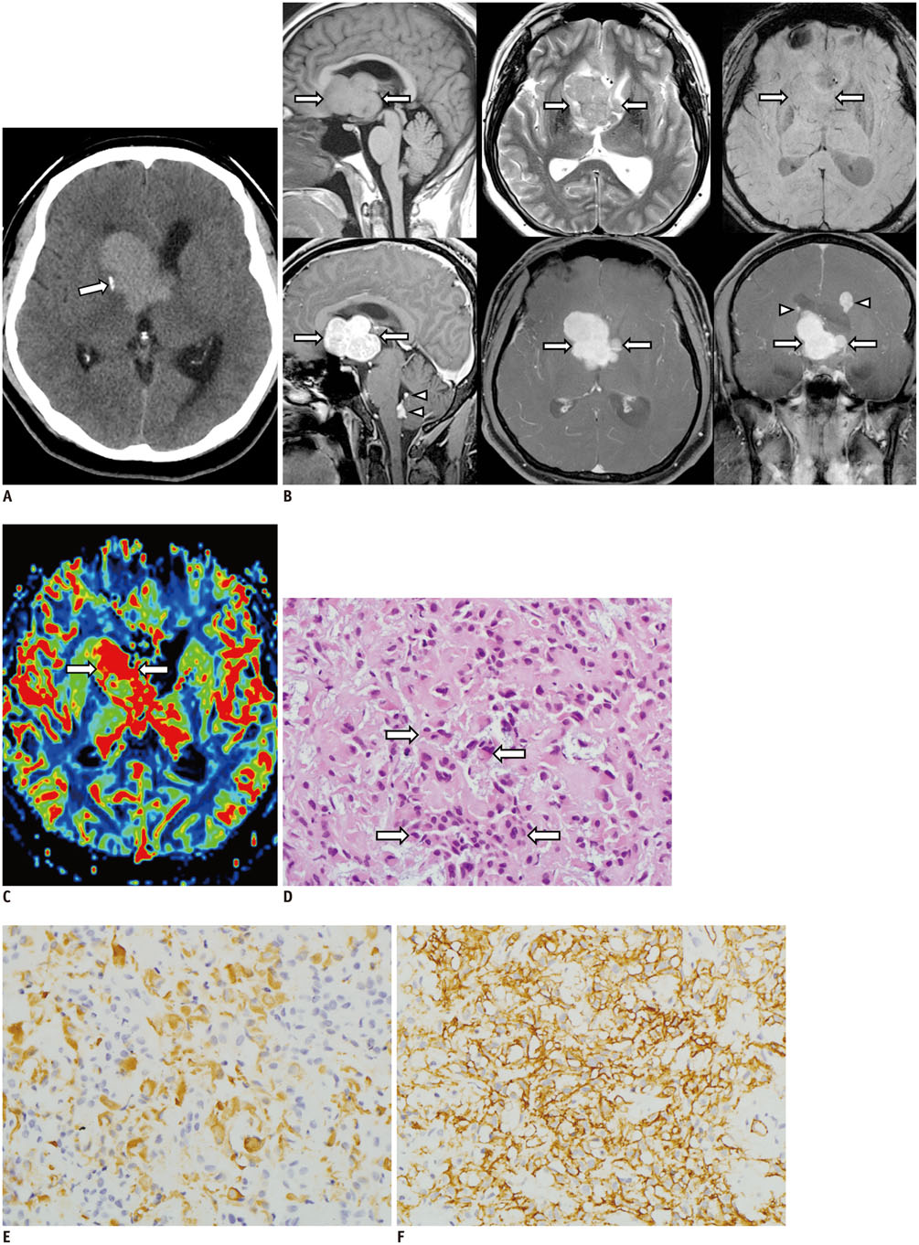
Fig. 1 Chordoid glioma in 34-year-old man. A. Axial unenhanced CT showing hyperattenuated mass compressing frontal horn of right lateral ventricle. Note small calcification (arrow) in periphery of mass. B. Sagittal T1-weighted (upper left panel) and axial T2-weighted (upper central panel) images showing isointense lobulated mass (arrows) relative to cerebral cortex in anterior third ventricle. Axial susceptibility-weighted image (upper right panel) showing no evidence of intratumoral hemorrhage. Sagittal (lower left panel), axial (lower central panel), and coronal (lower right panel) post-contrast T1-weighted images showing strong enhancing main tumor with lobulated margin (arrows) in anterior part of third ventricle and smaller enhancing masses (arrowheads) along wall of lateral ventricles and fourth ventricle. C. CBV map of perfusion MRI showing elevated CBV within tumor (arrows) in third ventricle. D. Photomicrograph of hematoxylin and eosin stained slide showing solid cellular components composed of clusters and cords of epithelioid tumor cells (arrows) within variable mucinous stroma (original magnification × 400). CBV = cerebral blood volume E, F. Photomicrographs of immunostained slides for GFAP (E) and CD 34 (F) showing diffuse and strong expression in tumor cells (dark yellow and brown colors) (original magnification × 400). GFAP = glial-fibrillary acid protein
Reference
-
1. Louis DN, Ohgaki H, Wiestler OD, Cavenee WK, Burger PC, Jouvet A, et al. The 2007 WHO classification of tumours of the central nervous system. Acta Neuropathol. 2007; 114:97–109.2. Tanboon J, Aurboonyawat T, Chawalparit O. A 29-year-old man with progressive short term memory loss. Brain Pathol. 2014; 24:103–106.3. Vanhauwaert DJ, Clement F, Van Dorpe J, Deruytter MJ. Chordoid glioma of the third ventricle. Acta Neurochir (Wien). 2008; 150:1183–1191.4. Kim JW, Kim JH, Choe G, Kim CY. Chordoid glioma: a case report of unusual location and neuroradiological characteristics. J Korean Neurosurg Soc. 2010; 48:62–65.5. Pasquier B, Péoc'h M, Morrison AL, Gay E, Pasquier D, Grand S, et al. Chordoid glioma of the third ventricle: a report of two new cases, with further evidence supporting an ependymal differentiation, and review of the literature. Am J Surg Pathol. 2002; 26:1330–1342.6. Agarwal S, Stevenson ME, Sughrue ME, Wartchow EP, Mierau GW, Fung KM. Features of intraventricular tanycytic ependymoma: report of a case and review of literature. Int J Clin Exp Pathol. 2014; 7:3399–3407.7. Ambekar S, Ranjan M, Prasad C, Santosh V, Somanna S. Fourth ventricular ependymoma with a distant intraventricular metastasis: report of a rare case. J Neurosci Rural Pract. 2013; 4:Suppl 1. S121–S124.8. Alvarez de Eulate-Beramendi S, Rigau V, Taillandier L, Duffau H. Delayed leptomeningeal and subependymal seeding after multiple surgeries for supratentorial diffuse low-grade gliomas in adults. J Neurosurg. 2014; 120:833–839.9. Grand S, Pasquier B, Gay E, Kremer S, Remy C, Le Bas JF. Chordoid glioma of the third ventricle: CT and MRI, including perfusion data. Neuroradiology. 2002; 44:842–846.10. Desouza RM, Bodi I, Thomas N, Marsh H, Crocker M. Chordoid glioma: ten years of a low-grade tumor with high morbidity. Skull Base. 2010; 20:125–138.
- Full Text Links
-
- Actions
-
Cited
- CITED
-
- Close
- Share
-
- Similar articles
-
- Chordoid Glioma of the Third Ventricle with Unusual MRI Features
- Chordoid Glioma: an Uncommon Tumor of the Third Ventricle
- Suprasellar Chordoid Glioma Combined with Rathke's Cleft Cyst: Case Report
- Chordoid Glioma : A Case Report of Unusual Location and Neuroradiological Characteristics
- Chordoid Glioma: A Case Report
