J Korean Ophthalmol Soc.
2012 Mar;53(3):478-481.
A Case of Congenital Idiopathic Microcoria
- Affiliations
-
- 1Department of Ophthalmology, Maryknoll Medical Center, Busan, Korea. daeyun115@hanmail.net
- 2Lee Eye Hospital, Busan, Korea.
Abstract
- PURPOSE
To report a case of congenital idiopathic microcoria corrected with pupilloplasty and amblyopia treatment.
CASE SUMMARY
A 4-year-old girl was referred for pupillary abnormality of the left eye. Her mother experienced no problems during gestation, and the patient was born at full term. On initial examination, visual acuity was 20/25 in the right eye and counting finger at 50 cm in the left eye. Slit lamp examination revealed that the left pupil was displaced superonasally. A band of fibrous tissue extended across the left pupil and there was no red reflex. There was very slight reaction to mydriatics. Using 23-gauge vitrectomy scissors, a pupilloplasty was performed, and the synechiae are removed. Postoperatively, the pupillary light reflex was brisk, and occlusion therapy was initiated. After 3 months, the visual acuity of the left eye improved to 20/30.
CONCLUSIONS
In our case, although microcoria was diagnosed at a relatively old age, the patient's clinical features were consistent with congenital idiopathic microcoria. Therefore, we diagnosed the patient with gradually progressed congenital idiopathic microcoria. In the case of typical congenital idiopathic microcoria, the red-reflex is absent at birth because of the small pupil. However, as in our case, it is possible that the pupil can contract gradually due to movement of the fibrous strand. The point must be considered in such patients.
MeSH Terms
Figure
-
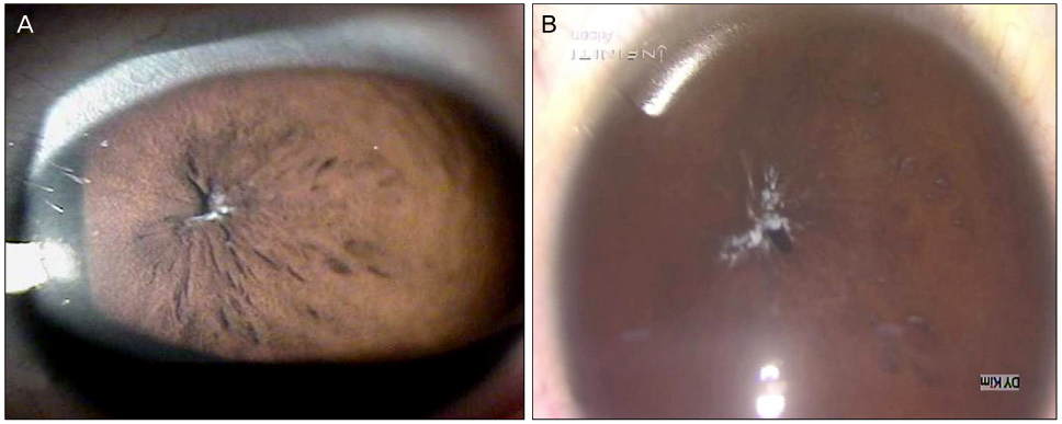
Figure 1 Preoperative anterior segment photograph shows a band of fibrous tissue extends across the pupil. The pupil is eccentric and the red reflex is absent. A pupil reaction to mydiratics is very slight. (A) Before topical infiltration of mydriatics. (B) After topical infiltration of mydriatics.
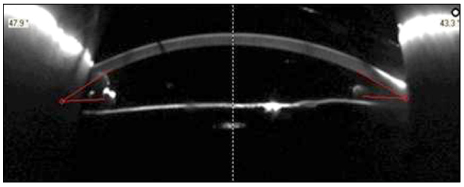
Figure 2 Scheimpflug image shows no pupillary opening.

Figure 3 B-scan image shows no vitreous opacity and any abnormalities.
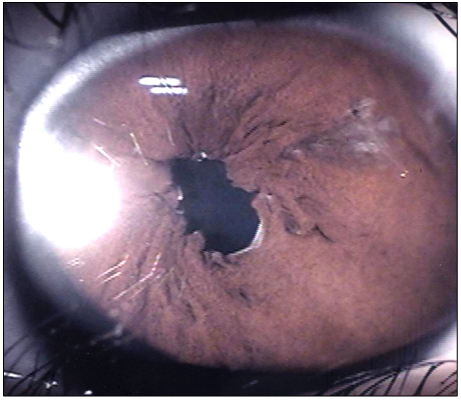
Figure 4 Postoperative anterior segment photography shows a widened pupil and irregular pupillary margin due to sphincterotomy.
Reference
-
1. Lambert SR, Amaya L, Taylor D. Congenital idiopathic microcoria. Am J Ophthalmol. 1988. 106:590–594.2. Weinstein JM, Zweifel TJ, Thompson HS. Congenital Horner's syndrome. Arch Ophthalmol. 1980. 98:1074–1078.3. Woodruff G, Buncic JR, Morin JD. Horner's syndrome in children. J Pediatr Ophthalmol Strabismus. 1988. 25:40–44.4. Mader TH, Wergeland FL Jr, Chismire KJ, Stephan MJ. Enlarged pupillary membranes. J Pediatr Ophthalmol Strabismus. 1988. 25:73–74.5. Holth S, Berner O. Congenital miosis of pinhole pupils owing to developmental faults of the dilatator muscle. Br J Ophthalmol. 1923. 7:401–419.6. Harms WA. Congenital eye defects. Congenital miosis associated with sex-linked congenital cataracts. J Kans Med Soc. 1977. 78:497–502.7. Aguirre Vila-Coro A, Mazow ML, Holladay JT, et al. Congenital idiopathic microcoria. Am J Ophthalmol. 1989. 107:439–440.8. Lim KH, Yu YS. Surgical management for persistent pupillary membrane with vitreous scissors. Korean J Ophthalmol. 1996. 10:124–126.
