J Korean Ophthalmol Soc.
2012 Feb;53(2):262-267.
Retinal Vascular Caliber Changes on OCT after Intravitreal Bevacizumab Injection in Diabetic Macular Edema
- Affiliations
-
- 1Department of Ophthalmology, Yonsei University College of Medicine, Seoul, Korea. semekim@yuhs.ac
- 2Department of Ophthalmology, CHA Bundang Medical Center, CHA University, Seongnam, Korea.
Abstract
- PURPOSE
To investigate retinal vascular caliber changes with Spectral Domain OCT after intravitreal bevacizumab injection in diabetic macular edema patients.
METHODS
Thirty-four eyes of 29 diabetic macular edema patients who were intravitreously injected with bevacizumab (1.25 g/0.05 ml) were studied. Twenty-four fellow eyes of 24 patients with bevacizumab injection were also recruited as a control group. We measured retinal vascular caliber a distance of 0.5-1 disc sizes from the disc margin with Spectral Domain OCT at baseline and 1-month and 3-month follow-up visits. Central macular thicknesses were also measured.
RESULTS
Over the 3 months of study, there was a significant reduction of arteriolar caliber compared to baseline (p = 0.024), There was significant reduction of venular caliber at 1 month (p = 0.001) and 3 months (p = 0.000) compared to baseline. Venular caliber reduction at the 3-month follow-up was significantly correlated with central macular thickness (R = 0.487, p = 0.003).
CONCLUSIONS
Retinal venular caliber reduction was significant at 1- and 3-month follow-up, and arteriolar caliber reduction was significant at 3-month follow-up compared to baseline. After 3 months of treatment, venular caliber reduction was correlated with reduction of central macular thickness.
MeSH Terms
Figure
-
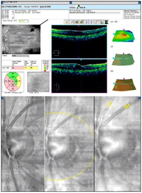
Figure 1 Measurement of vascular caliber. At 0.5-1 disc diameter from disc margin, vascular calibers are measured 3 times in arterioles and venules and the average is taken. Top) Main result of Cirrus HD-OCT. Fundus photograph is seen at the upper left of the screen (black arrow). Bottom left) Preoperative measurement of vascular caliber. Bottom center) Vascular calibers are measured at 0.5-1 disc size apart from disc margin. Bottom right) Measurement of vascular caliber at 3 months after intravitreal injection of bevacizumab.

Figure 2 Correlations of central macular thickness and retinal vascular caliber after intravitreal bevacizumab injection.
Reference
-
1. Ferris FL 3rd, Patz A. Macular edema. A complication of diabetic retinopathy. Surv Ophthalmol. 1984. 28:452–461.2. Kohner EM. Diabetic retinopathy. Clin Endocrinol Metab. 1977. 6:345–375.3. McDonald HR, Schatz H. Macular edema following panretinal photocoagulation. Retina. 1985. 5:5–10.4. McDonald HR, Schatz H. Visual loss following panretinal photocoagulation for proliferative diabetic retinopathy. Ophthalmology. 1985. 92:388–393.5. Seo JW, Park IW. Intravitreal bevacizumab for treatment of diabetic macular edema. Korean J Ophthalmol. 2009. 23:17–22.6. Velez-Montoya R, Fromow-Guerra J, Burgos O, et al. The effect of unilateral intravitreal bevacizumab (avastin), in the treatment of diffuse bilateral diabetic macular edema: a pilot study. Retina. 2009. 29:20–26.7. Chung EJ, Hong YT, Lee SC, et al. Prognostic factors for visual outcome after intravitreal bevacizumab for macular edema due to branch retinal vein occlusion. Graefes Arch Clin Exp Ophthalmol. 2008. 246:1241–1247.8. Joussen AM, Poulaki V, Qin W, et al. Retinal vascular endothelial growth factor induces intercellular adhesion molecule-1 and endothelial nitric oxide synthase expression and initiates early diabetic retinal leukocyte adhesion in vivo. Am J Pathol. 2002. 160:501–509.9. Ferrara N. Vascular endothelial growth factor: basic science and clinical progress. Endocr Rev. 2004. 25:581–611.10. Klein R, Sharrett AR, Klein BE, et al. Are retinal arteriolar abnormalities related to atherosclerosis?: The Atherosclerosis Risk in Communities Study. Arterioscler Thromb Vasc Biol. 2000. 20:1644–1650.11. Ikram MK, de Jong FJ, Vingerling JR, et al. Are retinal arteriolar or venular diameters associated with markers for cardiovascular disorders? The Rotterdam Study. Invest Ophthalmol Vis Sci. 2004. 45:2129–2134.12. Klein R, Klein BE, Knudtson M, et al. Are inflammatory factors related to retinal vessel caliber? The Beaver Dam Eye Study. Arch Ophthalmol. 2006. 124:87–94.13. Haritoglou C, Kook D, Neubauer A, et al. Intravitreal bevacizumab (Avastin) therapy for persistent diffuse diabetic macular edema. Retina. 2006. 26:999–1005.14. Arevalo JF, Fromow-Guerra J, Quiroz-Mercado H, et al. Primary intravitreal bevacizumab (Avastin) for diabetic macular edema: results from the Pan-American Collaborative Retina Study Group at 6-month follow-up. Ophthalmology. 2007. 114:743–750.15. Nguyen TT, Wang JJ, Sharrett AR, et al. Relationship of retinal vascular caliber with diabetes and retinopathy: the Multi-Ethnic Study of Atherosclerosis (MESA). Diabetes Care. 2008. 31:544–549.16. Fischer S, Renz D, Schaper W, Karliczek GF. In vitro effects of dexamethasone on hypoxia-induced hyperpermeability and expression of vascular endothelial growth factor. Eur J Pharmacol. 2001. 411:231–243.17. Wickremasinghe SS, Rogers SL, Gillies MC, et al. Retinal vascular caliber changes after intravitreal triamcinolone treatment for diabetic macular edema. Invest Ophthalmol Vis Sci. 2008. 49:4707–4711.18. Knudtson MD, Lee KE, Hubbard LD, et al. Revised formulas for summarizing retinal vessel diameters. Curr Eye Res. 2003. 27:143–149.19. Fukumura D, Gohongi T, Kadambi A, et al. Predominant role of endothelial nitric oxide synthase in vascular endothelial growth factor-induced angiogenesis and vascular permeability. Proc Natl Acad Sci U S A. 2001. 98:2604–2609.20. Goel N, Kumar V, Ghosh B. Ischemic maculopathy following intravitreal bevacizumab for refractory diabetic macular edema. Int Ophthalmol. 2011. 31:39–42.21. Lee SJ, Koh HJ. Enlargement of the foveal avascular zone in diabetic retinopathy after adjunctive intravitreal bevacizumab (avastin) with pars plana vitrectomy. J Ocul Pharmacol Ther. 2009. 25:173–174.22. Chen E, Hsu J, Park CH. Acute visual acuity loss following intravitreal bevacizumab for diabetic macular edema. Ophthalmic Surg Lasers Imaging. 2009. 40:68–70.
- Full Text Links
-
- Actions
-
Cited
- CITED
-
- Close
- Share
-
- Similar articles
-
- Macular Hole Formation after Intravitreal Injection of Bevacizumab for Diabetic Macular Edema
- Effects of Intravitreal Bevacizumab Injection in 3 Types of Macular Edema Secondary to Branch Retinal Vein Occlusion
- Comparison of Intravitreal Triamcinolone Versus Bevacizumab in Bilateral Diabetic Macular Edema by Optical Coherence Tomography (OCT) Patterns
- Electrophysiological and Morphological Changes After Intravitreal Bevacizumab Injection with Macular Edema or Choroidal Neovascularization
- The Effects of Intravitreal Bevacizumab Injection According to the Type of Diabetic Macular Edema
