Infected Paratracheal Air Cyst: A Case Report
- Affiliations
-
- 1Department of Radiology, Soonchunhyang University College of Medicine, Cheonan Hospital, Cheonan, Korea. ssmri@schmc.ac.kr
- KMID: 2327370
- DOI: http://doi.org/10.3348/jksr.2016.75.1.68
Abstract
- An air-filled paratracheal cyst is a common radiological finding. It may be a congenital defect or an acquired lesion. "Acquired paratracheal cyst" is the term given to the acquired abnormalities, which usually arise in adults. They result from a weakness of the tracheal wall, and they may be caused by trauma, infection, high pressure injuries, long lasting tracheostomy, and obstructive tracheal disease. Majority of the paratracheal air cysts are asymptomatic and are discovered incidentally on radiological images. Also, the management is primarily conservative treatment. Here, we report a case of an infected paratracheal air cyst on the right posterolateral wall of the trachea, which developed into an abscess and was visualized on follow-up multidetector computed tomography and was surgically removed due to persistent symptoms.
MeSH Terms
Figure
-
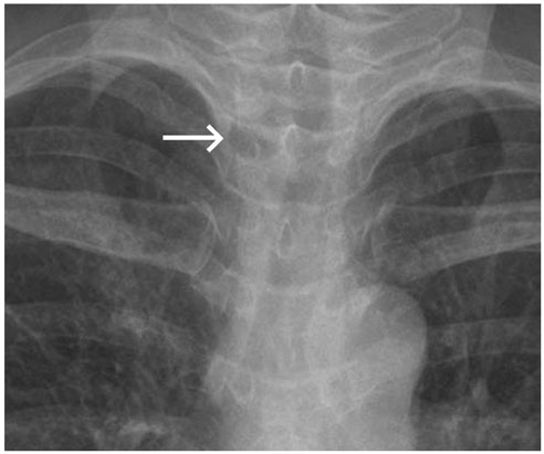
Fig. 1 Infected paratracheal air-cyst. A 53-year-old woman presented with a fever, sore throat, cough, sputum, and right neck pain for the past four days. Chest radiography demonstrates a round, radiolucent right paratracheal lesion (arrow) and right paratracheal stripe thickening.
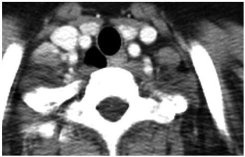
Fig. 2 Contrast-enhanced chest MDCT image obtained three years ago demonstrates an air-filled cystic lesion on the right side of the trachea without fat infiltration. MDCT = multidetector computed tomography
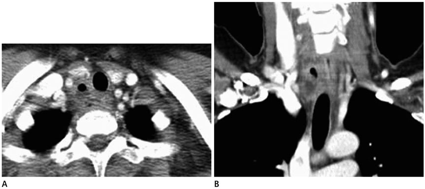
Fig. 3 Contrast-enhanced chest MDCT at present. Axial (A) and coronal reformatted (B) images demonstrate an air-filled and fluid-filled cystic lesion on the right side of the trachea, measuring about 2.5 × 2.2 × 1.3 cm in size, with an enhancing thick wall due to infection. MDCT = multidetector computed tomography
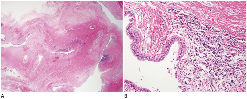
Fig. 4 A. Microscopically, the excised mass shows a multilocular cystic appearance in the low power view (hematoxylin and eosin, × 12.5). B. The lining cells are ciliated columnar epithelial cells. There is lymphoplasmacytic aggregation in the cystic walls (hematoxylin and eosin, × 200).
Cited by 1 articles
-
A Case of Retropharyngeal Abscess Resulting from Infected Paratracheal Cyst
Eun Soo Lee, Heon Soo Park, Sang Hyeon Kim, Dong Kun Lee
Korean J Otorhinolaryngol-Head Neck Surg. 2020;63(3):134-137. doi: 10.3342/kjorl-hns.2019.00549.
Reference
-
1. Cheng HM, Chang PY, Chiang KH, Huang HW, Lee CC. Prevalence and characteristics of paratracheal air cysts and their association with emphysema in a general population. Eur J Radiol. 2012; 81:2673–2677.2. Yazkan R, Ozpolat B, Firat H. Tracheocele; a case of rare clinical entity. Tuberk Toraks. 2008; 56:315–318.3. Kosehan D, Kayıhan A, Koktener A. Incidental right paratracheal air cyst: significance of 64-detector multislice CT in differential diagnosis. New J Med. 2011; 28:62–63.4. Boyaci N, Sen Dokumaci D, Karakas E, Yalcin F, Oney Kurnaz AG. Paratracheal air cysts: prevalence and relevance to pulmonary emphysema and bronchiectasis using thoracic multidetector CT. Diagn Interv Radiol. 2015; 21:42–46.5. S¸ahin N, Solak A, Genç B. A rare cause of odynophagia: infected tracheal diverticulum. Kulak Burun Bogaz Ihtis Derg. 2015; 25:126–130.6. Amaral CB, Silva S, Feijó S. Infected tracheal diverticulum: a rare association with alpha-1 antitrypsin deficiency. J Bras Pneumol. 2014; 40:669–672.7. Choi AR, Choi SH, Kim SW, Sung DW, Rha YH. A case of recurrent respiratory infection resulting from a congenital anomaly of the bronchial tree tracheal bronchus. Korean J Pediatr. 2008; 51:660–664.8. Teh BM, Hall C, Kleid S. Infected tracheocoele (acquired tracheal diverticulum): case report and literature review. J Laryngol Otol. 2011; 125:540–545.9. Polat AV, Elmali M, Aydin R, Ozbay A, Celenk C, Murat N. Paratracheal air cysts: prevalence and correlation with lung diseases using multi-detector CT. J Med Imaging Radiat Oncol. 2014; 58:144–148.10. Goo JM, Im JG, Ahn JM, Moon WK, Chung JW, Park JH, et al. Right paratracheal air cysts in the thoracic inlet: clinical and radiologic significance. AJR Am J Roentgenol. 1999; 173:65–70.
- Full Text Links
-
- Actions
-
Cited
- CITED
-
- Close
- Share
-
- Similar articles
-
- A Case of Retropharyngeal Abscess Resulting from Infected Paratracheal Cyst
- Paratracheal Air Cysts of Thoracic Inlet in Adults: CT Findings
- A Case of Paratracheal Air Cyst Mimicking an Upper Esophageal Diverticulum
- A Case of Bronchogenic Cyst with Infection of Paragonimus Westermani
- Paratracheal Air Cysts: Sonographic Findings in Two Cases
