CT Findings of Solitary Fibromatosis in the Colon: A Case Report
- Affiliations
-
- 1Department of Radiology, Myongji Hospital, Seonam University College of Medicine, Goyang, Korea. jhlim@mjh.or.kr
- 2Department of Pathology, Myongji Hospital, Seonam University College of Medicine, Goyang, Korea.
- KMID: 2327361
- DOI: http://doi.org/10.3348/jksr.2016.75.1.12
Abstract
- Fibromatosis is a rare benign neoplasm that appears as a sporadic lesion or is found in patients with familial adenomatous polyposis. Fewer than 7 cases of intraabdominal solitary fibromatosis arising from the colon have been reported in the English literature. This small number of reported cases may be not only because of the low incidence of the disease but also because of the difficulty in making proper diagnosis. We present here a case of histologically confirmed intraabdominal solitary fibromatosis arising from the colon, with an emphasis on computed tomography findings.
MeSH Terms
Figure
-
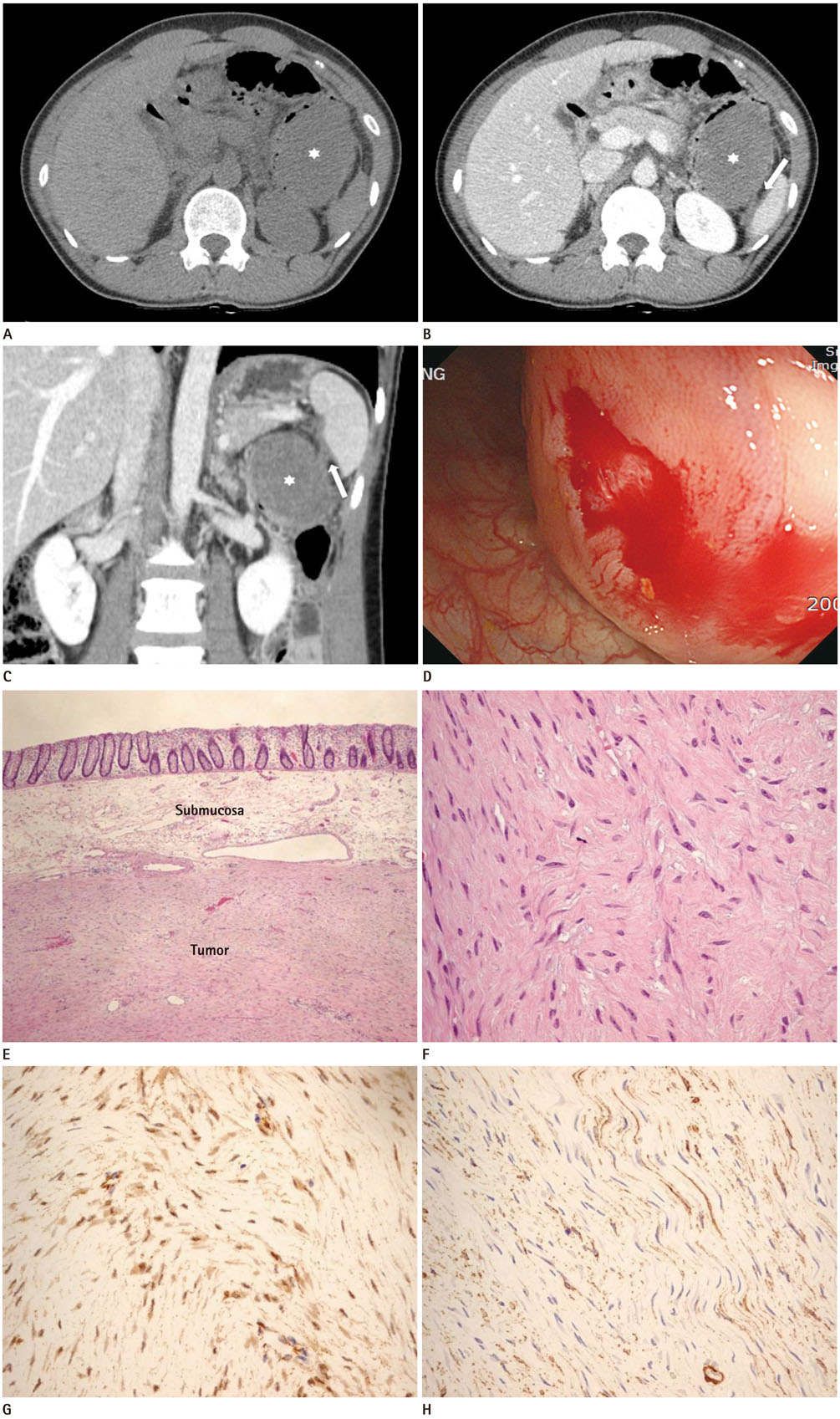
Fig. 1 A 43-year-old female with intraabdominal solitary fibromatosis of the colon. A. Axial image in the pre-contrast phase shows a 7 × 5 × 7 cm well-circumscribed homogeneous ovoid-shaped mass (asterisk) in the splenic flexure of the colon. B, C. Axial (B) and coronal (C) reformatted contrast-enhanced CT images in the portal venous phase show the mildly delayed enhancement of the mass (asterisks). There is fat infiltration and edematous change in the greater omentum, mesentery, and splenic hilum (arrows). D. Colonoscopy shows a 5 cm sized mass protruding into the lumen, covered with normal mucosa at the splenic flexure of the colon. E, F. Microscopic photomicrograph demonstrates that the tumor involves the submucosa of the colon (E, hematoxylin and eosin stain, × 40), and it is composed of fascicles of spindle cells with bland nuclear features and finely collagenous stroma (F, hematoxylin and eosin stain, × 400). G, H. Immunohistochemical stains (× 400) show immunopositivity for β-catenin (G) with nuclear accumulation and smooth muscle actin (H).
Reference
-
1. George V, Tammisetti VS, Surabhi VR, Shanbhogue AK. Chronic fibrosing conditions in abdominal imaging. Radiographics. 2013; 33:1053–1080.2. Eriguchi N, Aoyagi S, Okuda K, Hara M, Tamae T, Kanazawa N, et al. A case of Turner's syndrome complicated with desmoid tumor of the transverse colon. Kurume Med J. 1999; 46:181–184.3. Zhu Z, Li F, Zhuang H, Yan J, Wu C, Cheng W. FDG PET/CT detection of intussusception caused by aggressive fibromatosis. Clin Nucl Med. 2010; 35:370–373.4. Makis W, Ciarallo A, Abikhzer G, Stern J, Laufer J. Desmoid tumour (aggressive fibromatosis) of the colon mimics malignancy on dual time-point 18F-FDG PET/CT imaging. Br J Radiol. 2012; 85:e37–e40.5. Srigley JR, Mancer K. Solitary intestinal fibromatosis with perinatal bowel obstruction. Pediatr Pathol. 1984; 2:249–258.6. Al-Salem AH, Al-Hayek R, Qureshi SS. Solitary intestinal fibromatosis: a rare cause of intestinal perforation in neonates. Pediatr Surg Int. 1997; 12:437–440.7. Lacson AG, Harmel R, Neal MR, Welty P. Pathological case of the month. Solitary intestinal fibromatosis. Arch Pediatr Adolesc Med. 2000; 154:203–220.8. Numanoglu A, Davies J, Millar AJ, Rode H. Congenital solitary intestinal fibromatosis. Eur J Pediatr Surg. 2002; 12:337–340.9. Levy AD, Rimola J, Mehrotra AK, Sobin LH. From the archives of the AFIP: benign fibrous tumors and tumorlike lesions of the mesentery: radiologic-pathologic correlation. Radiographics. 2006; 26:245–264.10. Shinagare AB, Ramaiya NH, Jagannathan JP, Krajewski KM, Giardino AA, Butrynski JE, et al. A to Z of desmoid tumors. AJR Am J Roentgenol. 2011; 197:W1008–W1014.
- Full Text Links
-
- Actions
-
Cited
- CITED
-
- Close
- Share
-
- Similar articles
-
- Mesenteric Fibromatosis Representing as a Colo-Colic Intussusception Mimicking the Ascending Colon Malignant Tumor with CT and 18F-Fluorodeoxyglucose Positron Emission Tomography/CT Findings: A Case Report
- Mesenteric Fibromatosis with Spontaneous Cystic Degeneration: A Case Report with US and CT Findings
- Mesenteric Fibromatosis Causing Ureteral Stenosis
- Radiologic findings of mediastinal fibromatosis
- Desmoid type fibromatosis of the distal pancreas: A case report
