Two Cases of Caseous Calcification of the Mitral Annulus
- Affiliations
-
- 1Department of Medicine, Samsung Medical Center, Sungkyunkwan University School of Medicine, Seoul, Korea. parksmc@gmail.com
- KMID: 2297955
- DOI: http://doi.org/10.4070/kcj.2009.39.2.82
Abstract
- Caseous mitral annular calcification describes a heavy calcification of the mitral annulus with central liquefaction. This rare variant of mitral annular calcification may resemble an intracardiac tumor, abscess, vegetation, or thrombus, a resemblance that often leads to unnecessary surgery. Typical echocardiographic findings include a large, round, bright echogenic mass with a central echolucent area. It is known to have a benign clinical outcome, and it is thus managed conservatively. Because this entity is not well known and has only rarely been described, we report two cases of mitral annular calcification encountered at our institution. The first patient was an elderly woman with exertional dyspnea who was found to have a solitary pulmonary nodule on plain chest radiography. This was determined to represent caseous calcification of the mitral annulus. The other patient was an elderly woman who had a history of cerebral embolic infarction. She did not have an intracardiac thrombus, but she did have caseous mitral annular calcification. Both patients were managed conservatively.
Keyword
MeSH Terms
Figure
-
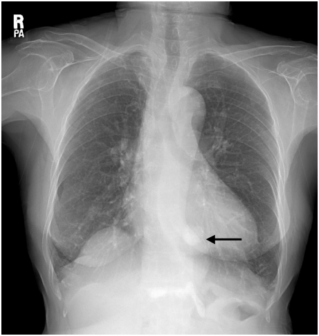
Fig. 1 Plain chest radiograph (posteroanterior projection) shows a 23 mm-sized nodule in the left lower lung zone, retrocardiac area (arrow).
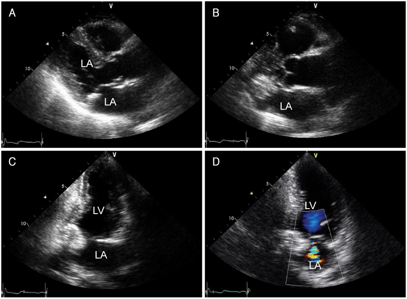
Fig. 2 Echocardiograph of case 1. A: parasternal long axis view shows a mitral annular calcification. B: zoomed image of the mitral valve shows a large, spherical echogenic mass with a smooth border. C: apical 2-chamber view of this calcified mass has a central echolucent area. D: color Doppler image shows no vascularity within the mass.
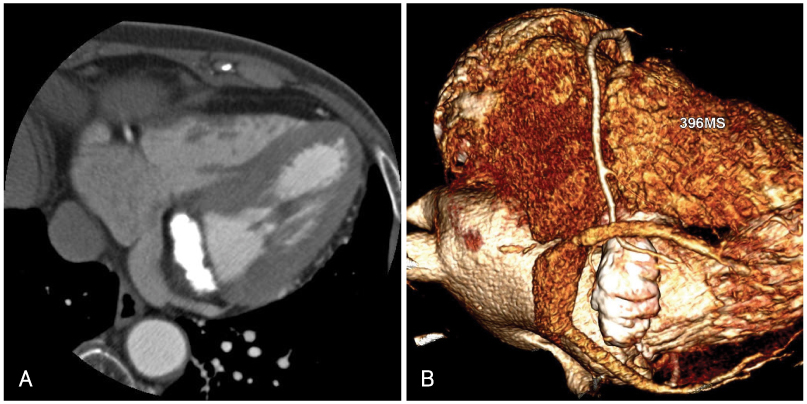
Fig. 3 Cardiac computed tomography. In the contrast-enhanced axial (A) and reconstructed volume rendering images (B), a dense calcified mass in the mitral annulus measuring 3.6×16×18 mm is shown. The coronary arteries are normal.
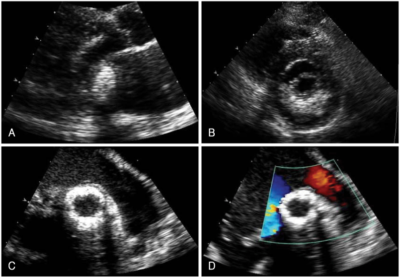
Fig. 4 Echocardiograph of case 2. Transthoracic echocardiography (A and B) and transesophageal echocardiography (C and D). A: parasternal long axis view of a spherical bright echogenic mass in the mitral annular area. B: parasternal short axis view of echogenic material around the mitral annular area. C: egg-shell like mass. D: color Doppler image shows mild mitral regurgitation and no significant flow communication between the central echolucent area and the cardiac chamber.
Reference
-
1. Zipes DP, Libby P, Bonow RO, Braunwald E. Braunwald's Heart Disease: A Textbook of Cardiovascular Medicine. 2005. 7 ed. Philadelphia: W.B. Saunders.2. Harpaz D, Auerbach I, Vered Z, Motro M, Tobar A, Rosenblatt S. Caseous calcification of the mitral annulus: a neglected, unrecognized diagnosis. J Am Soc Echocardiogr. 2001. 14:825–831.3. Teja K, Gibson RS, Nolan SP. Atrial extension of mitral annular calcification mimicking intracardiac tumor. Clin Cardiol. 1987. 10:546–548.4. Kato M, Nakatani S, Okazaki H, Tagusari O, Kitakaze M. Unusual appearance of mitral annular calcification mimicking intracardiac tumor prompting early surgery. Cardiology. 2006. 106:164–166.5. de Very EA, Scholte AJ, Krauss XH, et al. Intracardiac pseudotumor caused by mitral annular calcification. Eur J Echocardiogr. 2006. 7:62–66.6. Poh KK, Wood MJ, Cury RC. Prominent posterior mitral annular calcification causing embolic stroke and mimicking left atrial fibroma. Eur Heart J. 2007. 28:2216.7. Kronzon I, Winer HE, Cohen ML. Sterile, caseous mitral anular abscess. J Am Coll Cardiol. 1983. 2:186–190.8. Gilbert HM, Grodman R, Chung MH, Hartman G, Krieger KH, Hartman BJ. Sterile, caseous mitral valve "abscess" mimicking infective endocarditis. Clin Infect Dis. 1997. 24:1015–1016.9. Stone E, Cohn D, Deal C, Pollock C. Calcific atrial mass in end-stage renal failure. Nephrol Dial Transplant. 1997. 12:807–810.10. Gramenzi S, Mazzola AA, Tagliaferri B, et al. Caseous calcification of the mitral annulus: unusual case of spontaneous resolution. Echocardiography. 2005. 22:510–513.
- Full Text Links
-
- Actions
-
Cited
- CITED
-
- Close
- Share
-
- Similar articles
-
- The Predictors of Mitral Regurgitation in Percutaneous Mital Commussurotomy Using Inoue Balloon
- Mitral Valve Repair for Barlow’s Disease with Mitral Annular and Subvalvular Calcification: A Case Report
- Mitral Annulus Velocity Measured by Pulsed Wave Doppler Tissue Imaging in Healthy Korean People
- Assessment of Diastolic Function Using Mitral Annulus Velocity by Doppler Tissue Velocity in the Patients with Left Ventricular Hypertrophy
- Does The Mitral Annulus Shrink or Enlarge During Systole? A Real-Time 3D Echocardiography Study
