Ann Rehabil Med.
2012 Aug;36(4):565-568. 10.5535/arm.2012.36.4.565.
Retropharyngeal Abscess Initially Diagnosed by the Videofluoroscopic Swallowing Study
- Affiliations
-
- 1Department of Rehabilitation Medicine, Seoul National University College of Medicine, Seoul 110-744, Korea.
- 2Department of Rehabilitation Medicine, Seoul National University Boramae Medical Center, Seoul 156-707, Korea. ideale@empal.com
- KMID: 2266730
- DOI: http://doi.org/10.5535/arm.2012.36.4.565
Abstract
- In this article, we report a case where a videofluoroscopic swallowing study (VFSS) revealed the cause of a recently developed idiopathic dysphagia in a 66-year-old patient and enabled emergent treatment. The patient reported a 10-day history of fever, cough, sputum production, and progressive jaundice. He was then admitted to the hospital with suspicion of aspiration pneumonia. Despite treatment with antibiotics, fever and leukocytosis were persistent. As he also reported dysphagia, we performed the VFSS, which showed subglottic aspiration on all types of food and revealed a retropharyngeal mass causing mechanical compression. A contrast-enhanced computerized tomography (CT) of his neck was performed following the VFSS, which helped diagnose the mass as an extensive retropharyngeal abscess with mediastinitis. Following this diagnosis, emergent surgical incision and drainage was performed on the patient. Although the VFSS is primarily designed to evaluate swallowing function rather than to diagnose a disease, it can be used to reveal the primary medical cause of dysphagia while it studies the mechanical and structural abnormalities in the oropharyngeal and esophageal regions. This study also proposes that retropharyngeal abscess should be considered in the differential diagnosis of cases showing progressive dysphagia with fever. As confirmed through this work, the VFSS can function as a useful tool for detecting crucial diseases accompanying deglutition disorder.
MeSH Terms
Figure
-
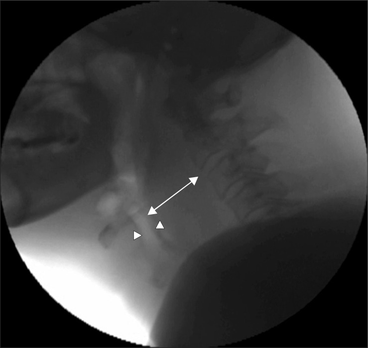
Fig. 1 Still image of the first VFSS. Retropharyngeal soft tissue was severely swollen (arrow) with a shifting airway (arrowhead) anteriorly.
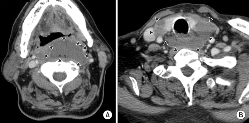
Fig. 2 Contrast-enhanced CT scan of the neck. Retropharyngeal abscess (arrowhead) was extended from upper pharynx (A) to the upper mediastinum (B).
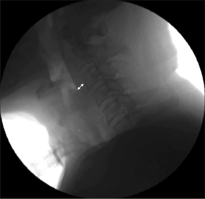
Fig. 3 Still image of the second VFSS. The thickness of the retropharyngeal soft tissue is normal (arrow).
Reference
-
1. Castell DO, Donner MW. Evaluation of dysphagia: a careful history is crucial. Dysphagia. 1987; 2:65–71. PMID: 3507297.
Article2. Wang LF, Kuo WR, Tsai SM, Huang KJ. Characterizations of life-threatening deep cervical space infections: a review of one hundred ninety-six cases. Am J Otolaryngol. 2003; 24:111–117. PMID: 12649826.
Article3. Ridder GJ, Technau-Ihling K, Sander A, Boedeker CC. Spectrum and management of deep neck space infections: an 8-year experience of 234 cases. Otolaryngol Head Neck Surg. 2005; 133:709–714. PMID: 16274797.4. Lazor JB, Cunningham MJ, Eavey RD, Weber AL. Comparison of computed tomography and surgical findings in deep neck infections. Otolaryngol Head Neck Surg. 1994; 111:746–750. PMID: 7991254.
Article5. Sichel JY, Dano I, Hocwald E, Biron A, Eliashar R. Nonsurgical management of parapharyngeal space infections: a prospective study. Laryngoscope. 2002; 112:906–910. PMID: 12150626.
Article6. Tannebaum RD. Adult retropharyngeal abscess: a case report and review of the literature. J Emerg Med. 1996; 14:147–158. PMID: 8740744.
Article
- Full Text Links
-
- Actions
-
Cited
- CITED
-
- Close
- Share
-
- Similar articles
-
- Dysphagia after Retropharyngeal Abscess Treated with Transcutaneous Injection of Botulinum Toxin at Cricopharyngeus Muscle
- A Case of Retropharyngeal Abscess and Mediastinal Abscess with Dyspnea
- Retropharyngeal Abscess Associated with Osteomyelitis of the Cervical Spine and an Epidural Abscess
- A case of Kawasaki disease with coexistence of a parapharyngeal abscess requiring incision and drainage
- Headache in the Retropharyngeal Abscess: A case report
