Anatomy and morphology of the nasopalatine canal using cone-beam computed tomography
- Affiliations
-
- 1Department of Oral Medicine and Radiology, Faculty of Dentistry, Jamia Millia Islamia University, New Delhi, India. arpitadoc@gmail.com
- 2Department of Oral Medicine and Radiology, S.D.M. College of Dental Sciences and Hospital, Dharwad, India.
- KMID: 2229612
- DOI: http://doi.org/10.5624/isd.2013.43.4.273
Abstract
- PURPOSE
This study was performed to evaluate the general anatomy and morphology of the nasopalatine canal using cone-beam computed tomography (CBCT) and to determine the human anatomic variability of the nasopalatine canal in relation to age and gender.
MATERIALS AND METHODS
The study included 100 subjects aged between 20 and 86 years who were divided into the following 3 groups: 1) 20-34 years old; 2) 35-49 years old; 3) > or =50 years old. The subjects were equally distributed between the genders. CBCT was performed using a standard exposure and patient positioning protocol. The data of the CBCT images were sliced in three dimensions. Image planes on the three axes (X, Y, and Z) were sequentially analyzed for the location, morphology and dimensions of the nasopalatine canal by two independent observers. The correlation of age and gender with all the variables was evaluated.
RESULTS
The present study did not reveal statistically significant differences in the number of openings at the nasal fossa; diameter of the nasal fossa openings; diameter of the incisive fossa; shape, curvature, and angulation of the canal as viewed in the sagittal sections; antero-posterior dimensions and length of the canal in the sagittal sections; or the level of division of the canal in the coronal plane by age. However, males and females showed significant differences in the length of the canal in the sagittal sections and level of the division of the canal in the coronal plane.
CONCLUSION
The present study highlighted important variability observed in the anatomy and morphology of the nasopalatine canal.
Figure
-
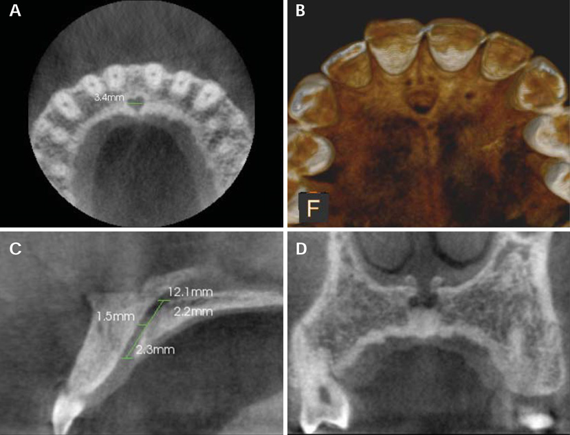
Fig. 1 The nasopalatine canal on CBCT images. A. An axial section at the level of the incisive fossa shows the medio-lateral diameter of the incisive fossa. B. A three-dimensional reconstructed image of the nasopalatine canal. C. A sagittal section shows the measurements of the length of the nasopalatine canal and the antero-posterior diameter of the canal at the nasal fossa, mid-level, and hard palate. D. A coronal section shows two openings of Stenson's foramina.
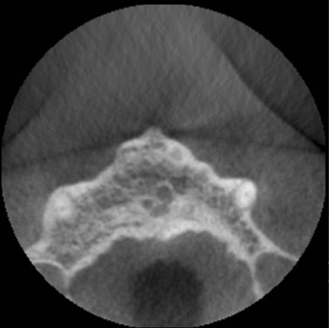
Fig. 2 A CBCT image shows 3 openings of the nasopalatine canal.
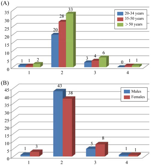
Fig. 3 Bar diagrams show the distribution of the number of openings at the nasal fossa. A. An age-wise distribution of the number of openings at the nasal fossa. B. A gender-wise distribution of the number of openings at the nasal fossa.

Fig. 4 CBCT images show the four shapes of the nasopalatine canal as seen on sagittal planes based on the labial and palatal walls of the nasopalatine canal. A. A cylindrical shape formed by parallel labial and palatal walls of the nasopalatine canal. B. A funnel shape formed by an increasing antero-posterior dimension of the nasopalatine canal from the nasal fossa to the hard palate. C. An hourglass shape in which the narrowest antero-posterior dimension of the NPC was at the mid-level compared to the dimensions at the nasal fossa and hard palate levels. D. A spindle shape in which the widest antero-posterior dimension of the nasopalatine canal was at the mid-level compared to the dimensions at the nasal fossa and hard palate levels.
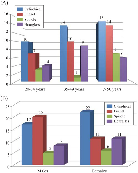
Fig. 5 Bar diagrams show the distribution of the subjects according to the shape of the canal. A. An age-wise distribution of the subjects according to the shape of the canal. B. A gender-wise distribution of the subjects according to the shape of the canal.
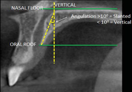
Fig. 6 A CBCT image in the sagittal plane shows the method of measurement for vertical and slanted nasopalatine canals.
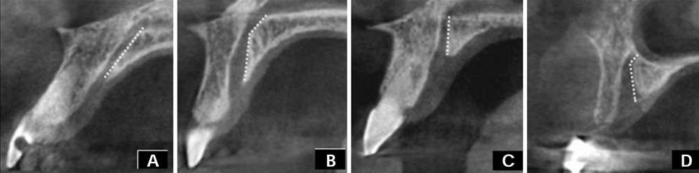
Fig. 7 CBCT images show the curvature of the nasopalatine canal as viewed in the sagittal plane. A. A slanted nasopalatine canal. B. A slanted-curved nasopalatine canal. C. A vertical nasopalatine canal. D. A vertical-curved nasopalatine canal.

Fig. 8 Bar diagrams show the distribution of the subjects according to the curvature of the canal. A. An age-wise distribution of the subjects according to the curvature of the canal. B. A gender-wise distribution of the subjects according to the curvature of the canal.
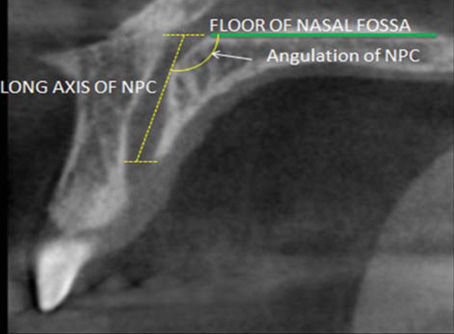
Fig. 9 A CBCT image in the sagittal plane shows the method of measurement for the angulation of the nasopalatine canal.
Cited by 1 articles
-
Unilateral maxillary central incisor root resorption after orthodontic treatment for Angle Class II, division 1 malocclusion with significant maxillary midline deviation: A possible correlation with root proximity to the incisive canal
Toshihiro Imamura, Shunsuke Uesugi, Takashi Ono,
Korean J Orthod. 2020;50(3):216-226. doi: 10.4041/kjod.2020.50.3.216.
Reference
-
1. Mardinger O, Namani-Sadan N, Chaushu G, Schwartz-Arad D. Morphologic changes of the nasopalatine canal related to dental implantation: a radiologic study in different degrees of absorbed maxillae. J Periodontol. 2008; 79:1659–1662.
Article2. Liang X, Jacobs R, Martens W, Hu Y, Adriaensens P, Quirynen M, et al. Macro- and micro-anatomical, histological and computed tomography scan characterization of the nasopalatine canal. J Clin Periodontol. 2009; 36:598–603.
Article3. Kraut RA, Boyden DK. Location of incisive canal in relation to central incisor implants. Implant Dent. 1998; 7:221–225.
Article4. Cavalcanti MG, Yang J, Ruprecht A, Vannier MW. Accurate linear measurements in the anterior maxilla using orthoradially reformatted spiral computed tomography. Dentomaxillofac Radiol. 1999; 28:137–140.
Article5. Artzi Z, Nemcovsky CE, Bitlitum I, Segal P. Displacement of the incisive foramen in conjunction with implant placement in the anterior maxilla without jeopardizing vitality of nasopalatine nerve and vessels: a novel surgical approach. Clin Oral Implants Res. 2000; 11:505–510.
Article6. Raghoebar GM, den Hartog L, Vissink A. Augmentation in proximity to the incisive foramen to allow placement of endosseous implants: a case series. J Oral Maxillofac Surg. 2010; 68:2267–2271.
Article7. Shear M, Speight P. Cysts of the oral and maxillofacial regions. 4th ed. Oxford: Blackwell;2007. p. 108–118.8. Bodin I, Isacsson G, Julin P. Cysts of the nasopalatine duct. Int J Oral Maxillofac Surg. 1986; 15:696–706.
Article9. Mraiwa N, Jacobs R, Van Cleynenbreugel J, Sanderink G, Schutyser F, Suetens P, et al. The nasopalatine canal revisited using 2D and 3D CT imaging. Dentomaxillofac Radiol. 2004; 33:396–402.
Article10. Sicher H. Anatomy and oral pathology. Oral Surg Oral Med Oral Pathol. 1962; 15:1264–1269.
Article11. Song WC, Jo DI, Lee JY, Kim JN, Hur MS, Hu KS, et al. Microanatomy of the incisive canal using three-dimensional reconstruction of microCT images: an ex vivo study. Oral Surg Oral Med Oral Pathol Oral Radiol Endod. 2009; 108:583–590.
Article12. Jacob S, Zelano B, Gungor A, Abbott D, Naclerio R, McClintock MK. Location and gross morphology of the nasopalatine duct in human adults. Arch Otolaryngol Head Neck Surg. 2000; 126:741–748.
Article
- Full Text Links
-
- Actions
-
Cited
- CITED
-
- Close
- Share
-
- Similar articles
-
- Observation of mandibular second molar roots and root canal morphology using dental cone-beam computed tomography
- Detection of maxillary second molar with two palatal roots using cone beam computed tomography: a case report
- Management of root canal perforation by using cone-beam computed tomography
- Three-dimensional imaging modalities in endodontics
- Cone-beam computed tomography analysis of root and canal morphology of mandibular premolars in a Spanish population
