A Case Report of Recurrent Subacute Stent Thrombosis After Repetitive Percutaneous Coronary Interventions
- Affiliations
-
- 1Division of Cardiology, Department of Internal Medicine, Soonchunhyang University College of Medicine, Bucheon, Korea. bluehartman@paran.com
- KMID: 2225853
- DOI: http://doi.org/10.4070/kcj.2008.38.2.122
Abstract
- Stent thrombosis (ST) is one of the major complications that occur in percutaneous coronary interventions (PCIs) with stents. Various factors have been attributed to the development of ST, and several strategies have been recommended for its management. We report the case of a patient suffering from recurrent subacute STs after recurrent PCIs. The patient was treated by coronary artery bypass graft (CABG).
Keyword
MeSH Terms
Figure
-
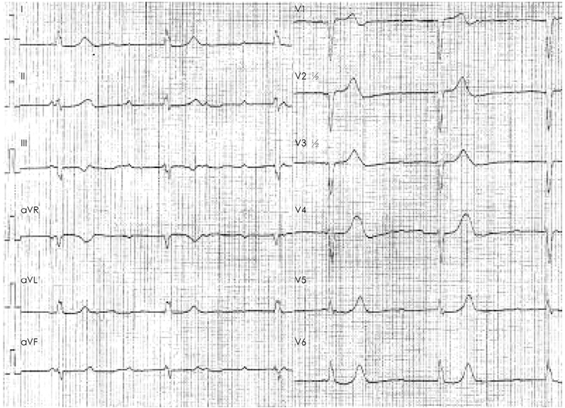
Fig. 1 ECG showed third degree atrioventricular block (data obtained from another hospital). ECG: electrocardiography.
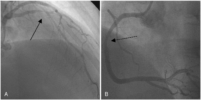
Fig. 2 Initial finding of coronary angiogram. CAG showed the culprit lesion (dark arrow) of the subtotally occluded proximal portion of the LAD with TIMI grade 3 flow (A) and 70% stenosis (dotted arrow) in the middle segment of the RCA (B). CAG: coronary angiography, LAD: left anterior descending artery, TIMI: thrombolysis in myocardial infarction, RCA: right coronary artery.
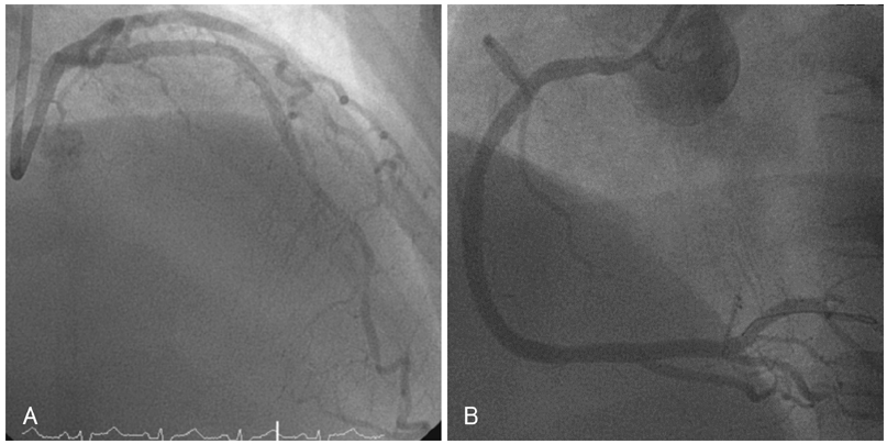
Fig. 3 CAG showed no residual stenosis in each lesion after 2 Cypher stents (Cordis/Johnson & Johnson, USA) were implanted in the lesions. A: post PCI angiography revealed well-deployed SES in mid portion of the LAD. B: post PCI angiography revealed well-deployed SES in mid portion of the RCA. CAG: coronary angiography, PCI: percutaneous coronary intervention.

Fig. 4 ECG showed ST segment elevation in leads V2-V4. ECG: electrocardiography.
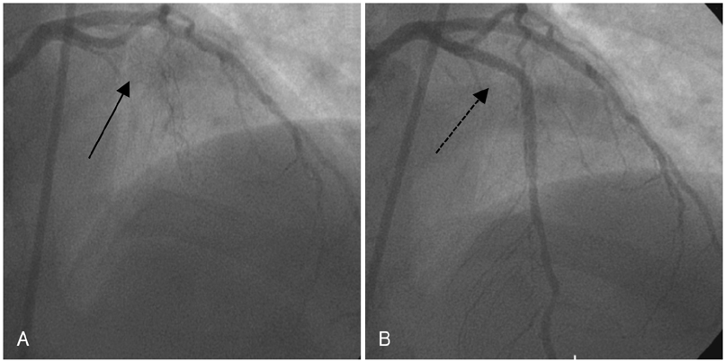
Fig. 5 Second findings of coronary angiogram. The second CAG showed total occlusion (dark arrow) in the previous stent-implanted area of the LAD (A) and restored lesion (dotted arrow) after successful PCI with ReoPro (abciximab) infusion (B). CAG: coronary angiography, LAD: left anterior descending artery, PCI: percutaneous coronary intervention.
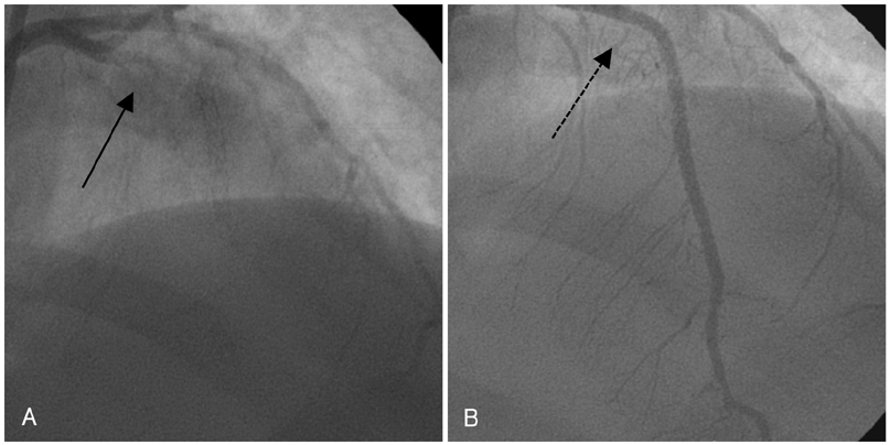
Fig. 6 The third finding of coronary angiogram. A, B: the third CAG showed total occlusion (dark arrow) of the pLAD and recanalized lesion (dotted arrow) after successful PCI. CAG: coronary angiography, pLAD: proximal left anterior descending artery, PCI: percutaneous coronary intervention.
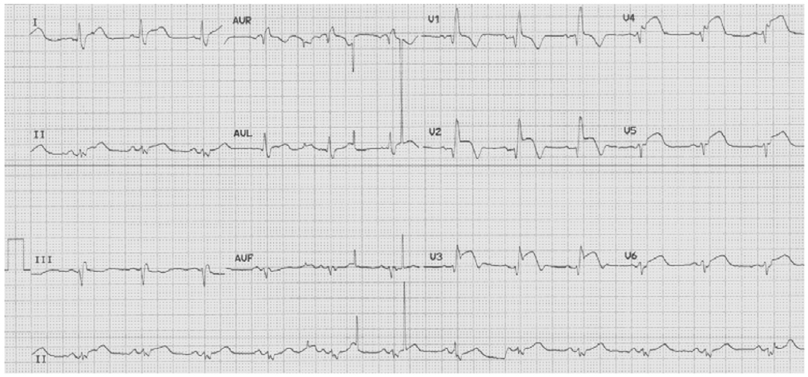
Fig. 7 ECG showed ST segment elevation in leads V2-V6. ECG: electrocardiography.
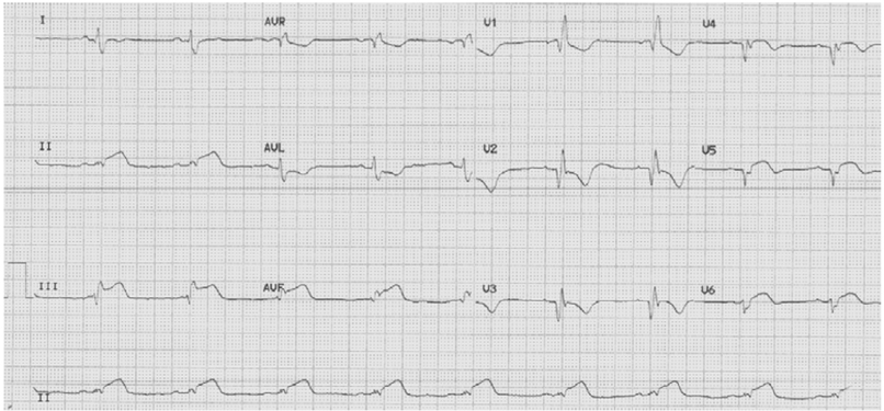
Fig. 8 ECG showed ST segment elevation in leads II, III and aVF. ECG: electrocardiography.
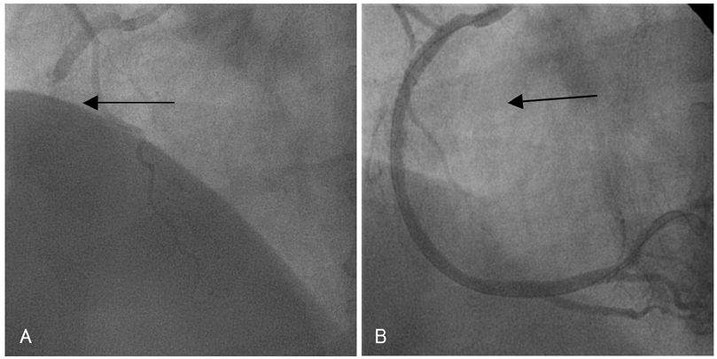
Fig. 9 The fifth finding of coronary angiogram. A, B: the fifth CAG showed total occlusion (dark arrow) in the earlier stent-implanted lesion of the RCA and recanalized lesion (dotted arrow) after successful PCI. CAG: coronary angiography, RCA: right coronary artery, PCI: percutaneous coronary intervention.
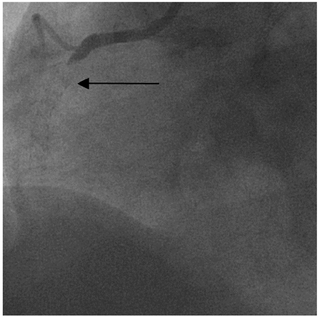
Fig. 10 The sixth CAG revealed exactly the same lesion (dark arrow) in the RCA 10 h after the PCI. CAG: coronary angiography, RCA: right coronary artery, PCI: percutaneous coronary intervention.
Reference
-
1. Kuchulakanti PK, Chu WW, Torguson R, et al. Correlates and long-term outcomes of angiographically proven stent thrombosis with sirolimus- and paclitaxel-eluting stents. Circulation. 2006. 113:1108–1113.2. Park DW, Park SW, Lee SW, et al. Frequency of coronary arterial late angiographic stent thrombosis (LAST) in the first six months: outcomes with drug-eluting stents versus bare metal stents. Am J Cardiol. 2007. 99:774–778.3. Mauri L, Hsieh WH, Massaro JM, Ho KK, D'Agostino R, Cutlip DE. Stent thombosis in randomized clinical trials of drung-eluting stents. N Engl J Med. 2007. 356:1020–1029.4. Stone GW, Moses JW, Ellis SG, et al. Safety and efficacy of sirolimus-and paclitaxel-eluting coronary stents. N Engl J Med. 2007. 356:998–1008.5. Park SH, Hong GR, Seo HS, Tahk SJ. Stent thrombosis after successful drug-eluting stent implantation. Korean Circ J. 2005. 35:163–171.6. Iakovou I, Schmidt T, Bonizzouni E, et al. Incidence, predictors, and outcome of thrombosis after successful implantation of drug eluting stents. JAMA. 2005. 293:2126–2130.7. Orford JL, Lennon R, Melby S, et al. Frequency and correlates of coronary stent thrombosis in the modern era: analysis of a single center registry. J Am Coll Cardiol. 2002. 40:1567–1572.8. Cutlip DE, Baim DS, Kalon K, et al. Stent thrombosis in the modern era. Circulation. 2001. 103:1967–1971.9. Uren NG, Schwarzacher SP, Metz JA, et al. Predictors and outcomes of stent thrombosis: an intravascular ultrasound registry. Eur Heart J. 2002. 23:124–132.10. Choi BR, Lee CW, Park SW. Late stent thrombosis associated with late stent malapposition after drug-eluting stenting: a case report. Korean Circ J. 2006. 36:472–475.11. Sucker C, Scheffold N, Cyran J, Ghodsizad A, Scharf RE, Zotz RB. No evidence for involvement of prothrombotic platelet receptor polymorphisms in acute coronary stent thrombosis. Int J Cardiol. 2008. [Epub ahead of print].12. Glowczynska R, Malek LA, Spiewak M, et al. Clinical, biochemical and genetical resistance to clopidogrel in a patient with the recurrent coronary stent thrombosis: a case report and review of the literature. Int J Cardiol. 2006. 111:326–328.13. Wenaweser P, Dorffler-Melly J, Imboden K, et al. Stent thrombosis is associated with an impared response to antiplatelet therapy. J Am Coll Cardiol. 2005. 45:1748–1752.
- Full Text Links
-
- Actions
-
Cited
- CITED
-
- Close
- Share
-
- Similar articles
-
- Acute Coronary Stent Thrombosis in Cancer Patients: A Case Series Report
- Subacute In-stent Thrombosis after Carotid Artery Stenting: A Case Report
- Recurrent Stent Thrombosis in Different Coronary Arteries
- Very Late Stent Thrombosis in Coronary Bare-Metal Stent Implantation: A Case Report
- A Case of Extremely Very Late Stent Thrombosis 8 Years after Implantation of Drug-Eluting Stent Observed by Intravascular Ultrasound
