J Korean Ophthalmol Soc.
2011 May;52(5):544-549. 10.3341/jkos.2011.52.5.544.
The Change in Corneal Endothelial Cell Density after Pars Plana Vitrectomy
- Affiliations
-
- 1Department of Ophthalmology, Soonchunhyang University College of Medicine, Seoul, Korea. wismile@unitel.co.kr
- 2Myeong Eye Clinic, Seoul, Korea.
- KMID: 2214573
- DOI: http://doi.org/10.3341/jkos.2011.52.5.544
Abstract
- PURPOSE
To assess the effect of pars plana vitrectomy and gas tamponade on corneal endothelial cell density.
METHODS
The corneal endothelial cell density in 145 eyes that underwent pars plana vitrectomy was examined with a noncontact specular microscope 3 months pre- and post-operation. The subjects were divided as follows: Group 1 (32 eyes) underwent pars plana vitrectomy, group 2 (26 eyes) underwent pars plana vitrectomy combined with gas tamponade, group 3 (34 eyes) underwent pars plana vitrectomy combined with phacoemulsification and group 4 (48 eyes) underwent pars plana vitrectomy combined with phacoemulsification and gas tamponade. The changes in corneal endothelial cell density between groups was compared.
RESULTS
The mean endothelial cell loss was more significant in group 2 than in group 1 (p = 0.012), and there was no difference between groups 3 and 4 (p = 0.063). However, after excluding 6 eyes that had blood in the center of the corneal endothelium as a result of being in the prone position following gas tamponade, the mean endothelial cell loss in group 2 was not less than in group 1. In eyes with blood in the corneal endothelium, endothelial cell loss significantly increased (p < 0.001).
CONCLUSIONS
The results of this study suggest that ophthalmic surgeons should attempt to carefully control bleeding and sufficiently irrigate the vitreous during pars plana vitrectomy combined with gas tamponade.
MeSH Terms
Figure
-
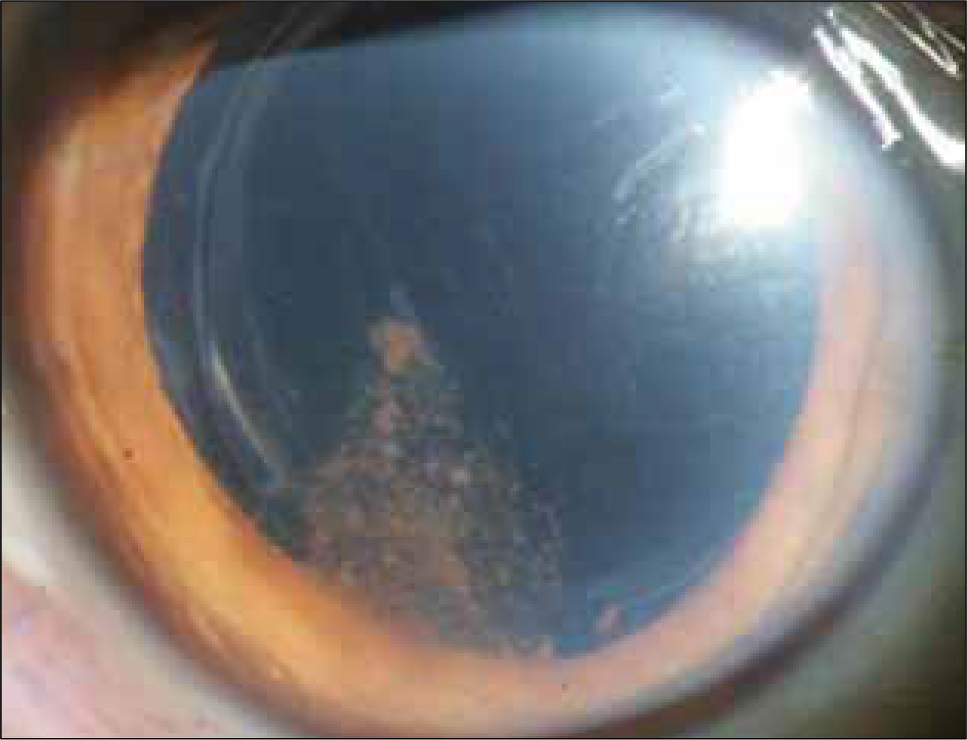
Figure 1. An anterior segment photograph of the patient who underwent pars plana vitrectomy, phacoemulsification and gas tamponade 2 weeks later. The component of blood is located between the pseudophakic lens and posterior capsule immediately, and then collected in the center of the corneal endothelium after being prone position.
Reference
-
References
1. Holzer MP, Tetz MR, Auffarth GU, et al. Effect of Healon5 and 4 other viscoelastic substances on intraocular pressure and endothelium after cataract surgery. J Cataract Refract Surg. 2001; 27:213–8.
Article2. Walkow T, Anders N, Klebe S. Endothelial cell loss after phacoemulsification: relation to preoperative and intraoperative parameters. J Cataract Refract Surg. 2000; 26:727–32.
Article3. Basti S, Aasuri MK, Reddy S, et al. Prospective evaluation of corneal endothelial cell loss after pediatric cataract surgery. J Cataract Refract Surg. 1998; 24:1469–73.
Article4. Baradaran-Rafii A, Rahmati-Kamel M, Eslani M, et al. Effect of hydrodynamic parameters on corneal endothelial cell loss after phacoemulsification. J Cataract Refract Surg. 2009; 35:732–7.
Article5. Jacobs PM, Cheng H, Price NC, et al. Endothelial cell loss after cataract surgery-the problem of interpretation. Trans Ophthalmol Soc U K. 1982; 102:291–3.6. Buettner H, Bourne WM. Effect of trans pars plana surgery on the corneal endothelium. Dev Ophthalmol. 1981; 2:28–34.7. Diddie KR, Schanzlin DJ. Specular microscopy in pars plana vitrectomy. Arch Ophthalmol. 1983; 101:408–9.
Article8. Friberg TR, Doran DL, Lazenby FL. The effect of vitreous and retinal surgery on corneal endothelial cell density. Ophthalmology. 1984; 91:1166–9.
Article9. Mitamura Y, Yamamoto S, Yamazaki S. Corneal endothelial cell loss in eyes undergoing lensectomy with and without anterior lens capsule removal combined with pars plana vitrectomy and gas tamponade. Retina. 2000; 20:59–62.
Article10. Agapitos PJ, Noel LP, Clarke WN. Traumatic hyphema in children. Ophthalmology. 1987; 94:1238–41.
Article11. Fischbarg J. Active and passive properties of the rabbit corneal endothelium. Exp Eye Res. 1973; 15:615–38.
Article12. Stiemke MM, Watsky MA, Kangas TA, Edelhauser HF. The establishment and maintenance of corneal transparency. Prog Retinal Eye Res. 1995; 14:109–40.13. Sugar J, Mitchelson J, Kraff M. The effect of phacoemulsification on corneal endothelial cell density. Arch Ophthalmol. 1978; 96:446–8.
Article14. Matsuda M, Miyake K, Inaba M. Long-term corneal endothelial changes after intraocular lens implantation. Am J Ophthalmol. 1988; 105:248–52.
Article15. Masket S. One year postoperative astigmatic comparison of sutured and unsutured 4.0 mm scleral pocket incisions. J Cataract Refract Surg. 1993; 19:453–6.
Article16. Mitamura Y, Takeuchi S, Kasai H, et al. Corneal endothelial cell damage associated with pars plana vitrectomy. Nippon Ganka Gakkai Zasshi. 1998; 102:59–63.17. Mittl RN, Koester CJ, Kates MR, Wilkes E. Endothelial cell counts following pars plana vitrectomy in pseudophakic and aphakic eyes. Ophthalmic Surg. 1989; 20:13–6.
Article18. Bourne RR, Minassian DC, Dart JK, et al. Effect of cataract surgery on the corneal endothelium: modern phacoemulsification compared with extracapsular cataract surgery. Ophthalmology. 2004; 111:679–85.19. Millá E, Vergés C, Ciprés M. Corneal endothelium evaluation after phacoemulsification with continuous anterior chamber infusion. Cornea. 2005; 24:278–82.
Article20. Green K, Cheeks L, Stewart DA, Norman BC. Intraocular gas effects on corneal endothelial permeability. Lens Eye Toxic Res. 1992; 9:85–91.21. Lee DA, Wilson MR, Yoshizumi MO, Hall M. The ocular effects of gases when injected into the anterior chamber of rabbit eyes. Arch Ophthalmol. 1991; 109:571–5.
Article22. Matsuda M, Tano Y, Inaba M, Manabe R. Corneal endothelial cell damage associated with intraocular gas tamponade during pars plana vitrectomy. Jpn J Ophthalmol. 1986; 30:324–9.23. Pong J, Lai J. Effect on corneal endothelial cell count of traumatic microhyphaema and hyphaema. Acta Ophthalmol. 2009; 87:559–61.
Article24. Messmer EP, Gottsch J, Font RL. Blood staining of the cornea: a histopathologic analysis of 16 cases. Cornea. 1984-1985; 3:205–12.
- Full Text Links
-
- Actions
-
Cited
- CITED
-
- Close
- Share
-
- Similar articles
-
- The Effects of Pars Plana Vitrectomy on the Corneal Endothelium
- Corneal Endothelial change following Par Plana Vitrectomy with Expanding Gas
- Long-term Changes of Endothelial Cell Density after Pars Plana Vitrectomy with Fragmentation
- Combined Clear Corneal Phacoemulsification and Pars Plana Vitrectomy
- The Alteration of Accommodative Power after Scleral Buckling and Pars Plana Vitrectomy
