J Korean Ophthalmol Soc.
2015 Oct;56(10):1552-1557. 10.3341/jkos.2015.56.10.1552.
The Relationship between the Nuclear Density Using Scheimpflug Imaging with Phacoemulsification Parameters
- Affiliations
-
- 1Department of Ophthalmology, Konyang University College of Medicine, Daejeon, Korea. kopupil@hanmail.net
- KMID: 2214414
- DOI: http://doi.org/10.3341/jkos.2015.56.10.1552
Abstract
- PURPOSE
To evaluate the correlation between nuclear density measured using the Pentacam(R) (Oculus Optikgerate GmbH, Wetzlar, Germany) Scheimpflug imaging system and phacoemulsification parameters.
METHODS
The nuclear density was measured using maximal nuclear density with ImageJ, Pentacam(R) nucleus staging (PNS), average value and maximal value in lens densitometry. Intraoperatively, effective phaco time (EPT) and balanced salt solution (BSS) used were noted and compared with the nuclear density calculation methods. As an index of corneal endothelial cells, the changes in cell density (CD) were compared with the nuclear density.
RESULTS
Regarding EPT, maximal nuclear density with ImageJ (r = 0.379, p < 0.01) and PNS (r = 0.367, p < 0.01) were correlated positively, but the other methods were not correlated. Concerning BSS used, maximal nuclear density with ImageJ (r = 0.279, p = 0.03) and PNS (r = 0.286, p = 0.04) were positively correlated, but the other methods were not correlated. The specular microscopy showed that as the nuclear density increased, the postoperative CD tended to decrease, but without statistical significance.
CONCLUSIONS
Preoperative nuclear density measurements using maximal nuclear density with ImageJ or PNS were correlated with phacoemulsification parameters.
Figure
-
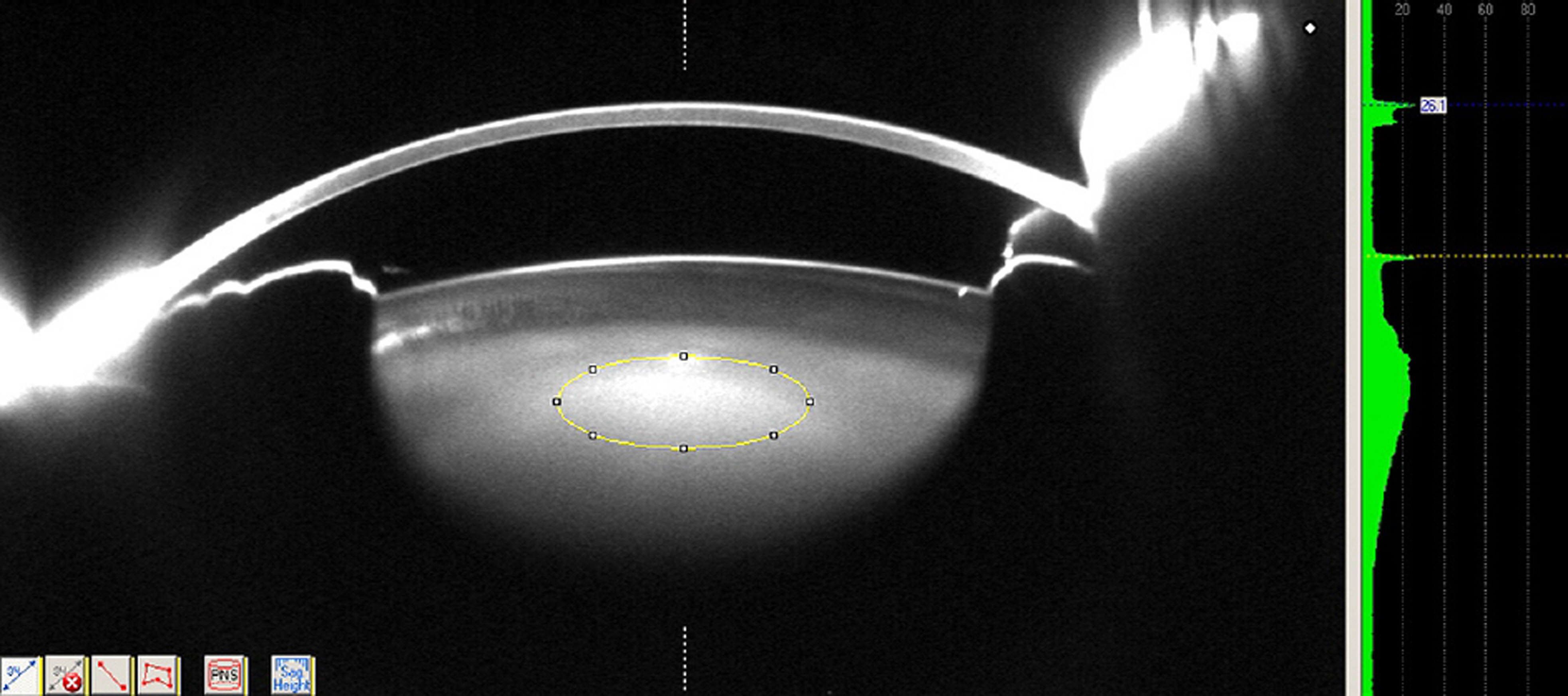
Figure 1. Scheimpflug image of the lens exported to ImageJ software for measuring the maximum nuclear lens density in the region of interest, indicated by the elliptical mask.
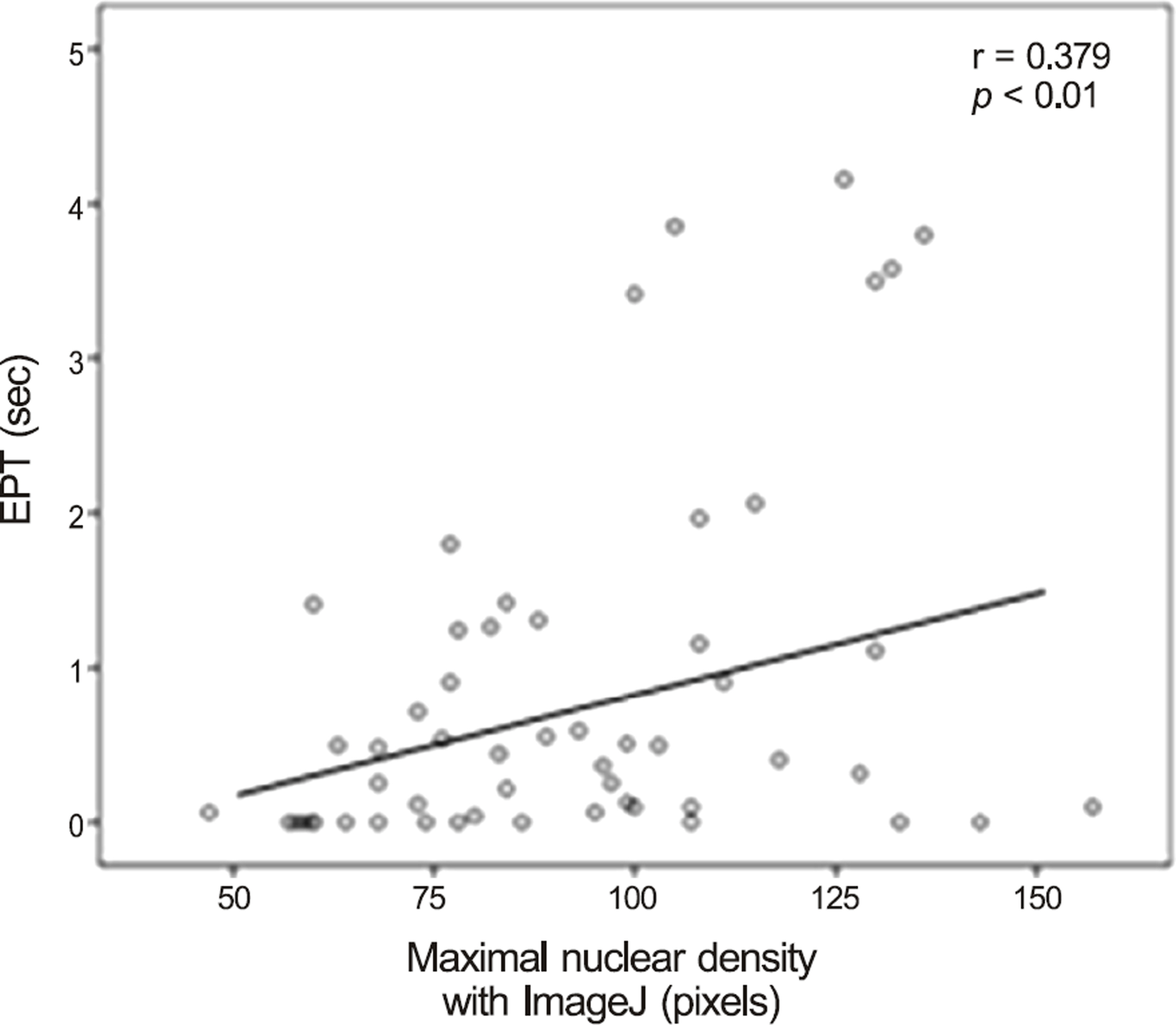
Figure 2. Scatter plot and relationship between maximal nu-clear density with ImageJ (pixels) and EPT (seconds). EPT = effective phaco time.
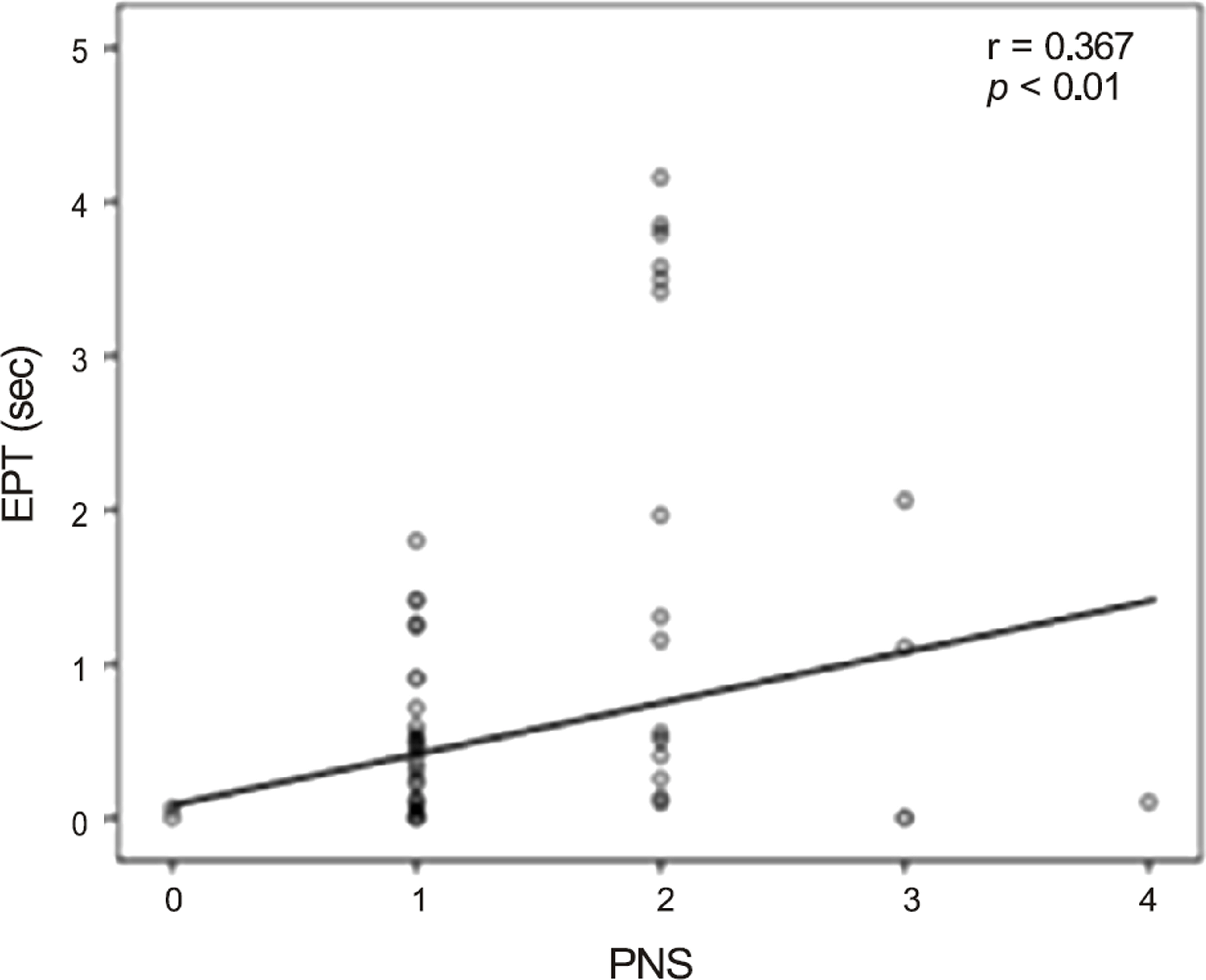
Figure 3. Scatter plot and relationship between PNS and EPT (seconds). PNS = Pentacam® nucleus staging; EPT = effec-tive phaco time.
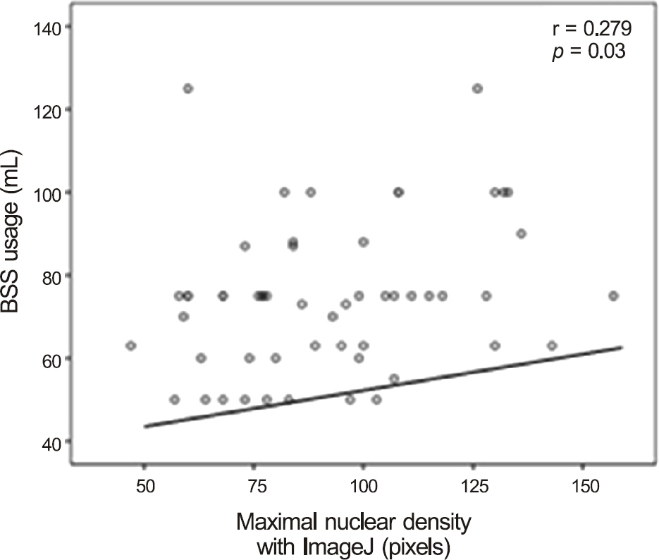
Figure 4. Scatter plot and relationship between maximal nu-clear density with ImageJ (pixels) and BSS usage (mL). BSS = balanced salt solution.
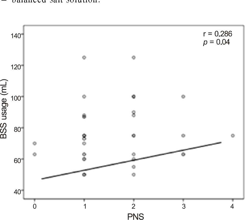
Figure 5. Scatter plot and relationship between PNS and BSS usage (mL). PNS = Pentacam® nucleus staging; BSS = balanced salt solution.
Reference
-
References
1. Paik HJ, Song HJ, Shyn KH. 2007 Survey for KSCRS mem-bers-current trends in cataract surgery in Korea. J Korean Ophthalmol Soc. 2009; 50:1624–31.2. Nixon DR. Preoperative cataract grading by Scheimpflug imaging and effect on operative fluidics and phacoemulsification energy. J Cataract Refract Surg. 2010; 36:242–6.
Article3. Kirwan JF, Venter L, Stulting AA, Murdoch IE. LOCS III examina-tion at the slit lamp, do settings matter? Ophthalmic Epidemiol. 2003; 10:259–66.4. Tan AC, Loon SC, Choi H, Thean L. Lens Opacities Classification System III: cataract grading variability between junior and senior staff at a Singapore hospital. J Cataract Refract Surg. 2008; 34:1948–52.
Article5. Chylack LT Jr, Wolfe JK, Singer DM. . The Lens Opacities Classification System III. The longitudinal study of Cataract Study Group. Arch Ophthalmol. 1993; 111:831–6.6. Grewal DS, Brar GS, Grewal SP. Correlation of nuclear cataract lens density using Scheimpflug images with Lens Opacities Classification System III and visual function. Ophthalmology. 2009; 116:1436–43.
Article7. Kim JS, Chung SH, Joo CK. Clinical application of a Scheimpflug system for lens density measurements in phacoemulsification. J Cataract Refract Surg. 2009; 35:1204–9.
Article8. Park CW, Kim GY, Kim HJ. . Objective clinical evaluation of ocular optical instrument according to the type of lens opacity. J Korean Ophthalmol Soc. 2014; 55:79–84.
Article9. Magalhães FP, Costa EF, Cariello AJ. . Comparative analysis of the nuclear lens opalescence by the Lens Opacities Classification System III with nuclear density values provided by Oculus Pentacam: a cross-section study using Pentacam Nucleus Staging software. Arq Bras Oftalmol. 2011; 74:110–3.
Article10. Gupta M, Ram J, Jain A. . Correlation of nuclear density using the Lens Opacity Classification System III versus Scheimpflug imaging with phacoemulsification parameters. J Cataract Refract Surg. 2013; 39:1818–23.
Article11. Kelman CD. The history and development of phacoemulsification. Int Ophthalmol Clin. 1994; 34:1–12.
Article12. Davison JA, Chylack LT. Clinical application of the lens opacities classification system III in the performance of phacoemulsification. J Cataract Refract Surg. 2003; 29:138–45.
Article13. Pei X, Bao Y, Chen Y, Li X. Correlation of lens density measured using the Pentacam Scheimpflug system with the Lens Opacities Classification System III grading score and visual acuity in age-re-lated nuclear cataract. Br J Ophthalmol. 2008; 92:1471–5.
Article14. Robman LD, McCarty CA, Garrett SK. . Comparison of clin-ical and digital assessment of nuclear optical density. Ophthalmic Res. 1999; 31:119–26.
Article15. Pirazzoli G, D’Eliseo D, Ziosi M, Acciarri R. Effects of phacoe-mulsification time on the corneal endothelium using phacofracture and phaco chop techniques. J Cataract Refract Surg. 1996; 22:967–9.
Article16. Hayashi K, Hayashi H, Nakao F, Hayashi F. Risk factors for cor-neal endothelial injury during phacoemulsification. J Cataract Refract Surg. 1996; 22:1079–84.
Article17. Bourne RR, Minassian DC, Dart JK. . Effect of cataract sur-gery on the corneal endothelium: modern phacoemulsification compared with extracapsular cataract surgery. Ophthalmology. 2004; 111:679–85.18. Walkow T, Anders N, Klebe S. Endothelial cell loss after phacoe-mulsification: relation to preoperative and intraoperative parameters. J Cataract Refract Surg. 2000; 26:727–32.
Article
- Full Text Links
-
- Actions
-
Cited
- CITED
-
- Close
- Share
-
- Similar articles
-
- The Relationship with the Density of Lens Nucleus and Phacoemulsification Time
- The Effect of Phacoemulsification with Oscillation Device on the Cornea and Lens Opcatiy
- The Relationship of the Lens Density with the Lens Thickness and the Anterior Chamber Depth
- Comparison of Phacoemulsification Time: Oscillation Device versus Conventional Device
- Periodic Analysis of Senile Cataract with EAS-1000 Scheimpflug Photography
