Surgically Induced Posterior Corneal Astigmatism in 2.2 mm Microcoaxial Cataract Surgery Versus 2.85 mm Coaxial Conventional Cataract Surgery
- Affiliations
-
- 1Department of Ophthalmology, The Catholic University of Korea College of Medicine, Seoul, Korea. sara514@catholic.ac.kr
- KMID: 2214412
- DOI: http://doi.org/10.3341/jkos.2015.56.10.1534
Abstract
- PURPOSE
To compare the surgically induced posterior astigmatism of microcoaxial cataract surgery using a 2.2 mm incision and conventional cataract surgery using a 2.85 mm incision.
METHODS
This study included 56 eyes that underwent phacoemulsification and intraocular lens insertion. Subjects were divided into 2 groups: 26 eyes receiving a microcoaxial cataract surgery using a 2.2 mm incision (MCCS group) and 30 eyes receiving a conventional cataract surgery using a 2.85 mm incision (CCS group). Anterior, posterior and total corneal astigmatism was measured. The surgically induced anterior astigmatism, surgically induced posterior astigmatism and surgically induced total astigmatism were compared between MCCS and CCS groups. Corneal astigmatism was measured using a Pentacam(R) device (Oculus, Wetzlar, Germany), uncorrected visual acuity, best corrected visual acuity and corneal aberrations of front and rear side was measured preoperatively and at 1 day, 1 month and 2 months postoperatively.
RESULTS
There was no difference in surgically induced posterior astigmatism between CCS and MCCS groups, however, surgically induced anterior astigmatism and surgically induced total astigmatism were significantly lower in the MCCS group than in the CCS group (p = 0.005 and, p = 0.036, respectively). There was a significant positive linear correlation between surgically induced posterior astigmatism and surgically induced total astigmatism in the CCS group (p = 0.01, r = 0.563). There was also a significant positive linear correlation between surgically induced anterior astigmatism and surgically induced total astigmatism in both CCS and MCCS groups (CCS group: p = 0.00, r = 0.855; MCCS group: p = 0.039, r = 0.407).
CONCLUSIONS
There was no significant difference in the surgically induced posterior astigmatism between the MCCS and CCS groups. However, surgically induced posterior astigmatism significantly affected surgically induced total astigmatism in the CCS group but not in the MCCS group. Considering both anterior and posterior astigmatism of the cornea, microcoaxial cataract surgery using a 2.2 mm incision affects surgically induced total astigmatism less than conventional cataract surgery.
Keyword
Figure
-
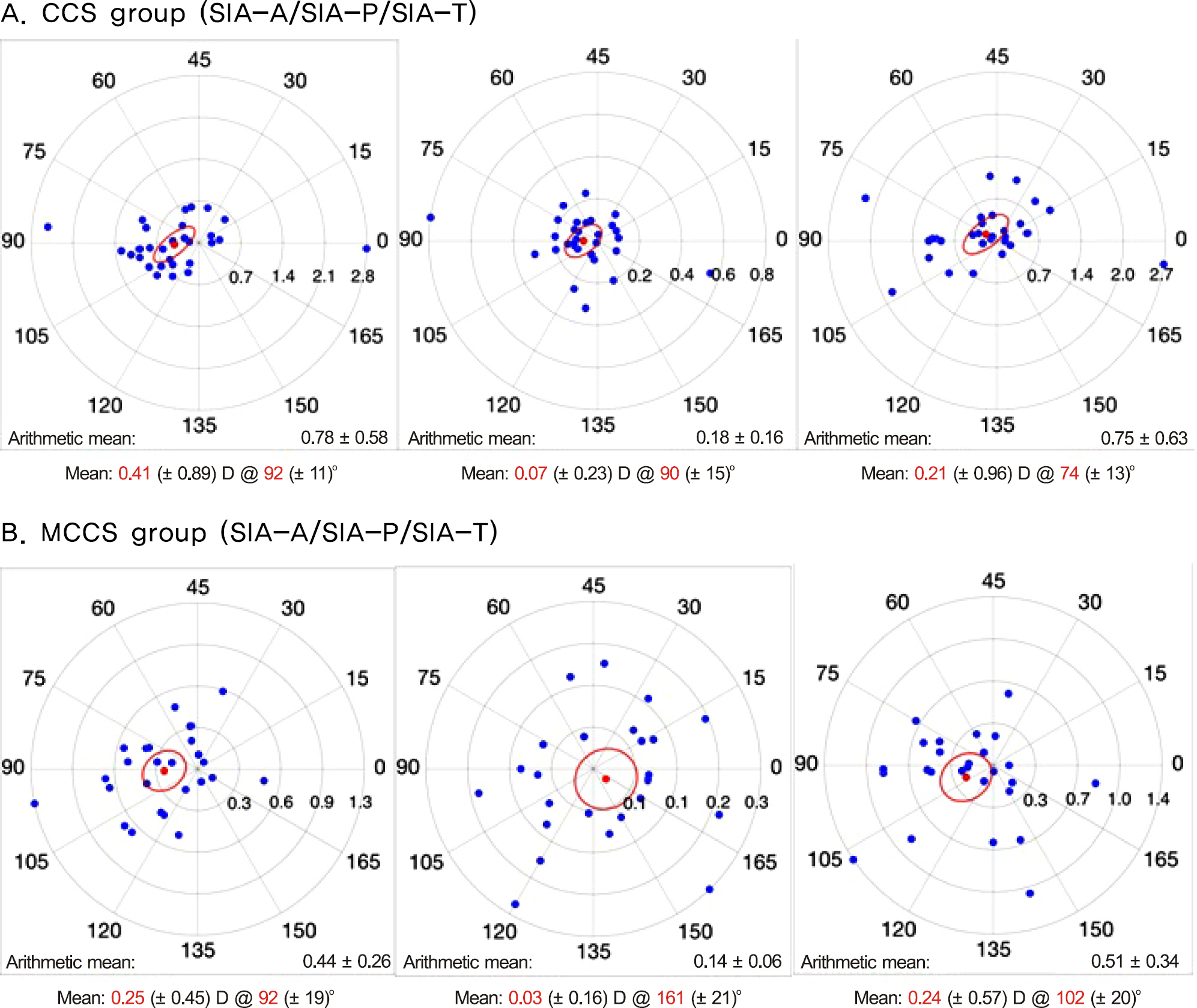
Figure 1. Double-angle plots of surgically induced astigmatism (SIA) using Pentacam® (Oculus, Wetzlar, Germany) at 2 months af-ter surgery, in CCS group (A) and MCCS group (B). CCS = conventional cataract surgery; MCCS = microcoaxial cataract sur-gery; SIA-A = surgically induced astigmatism-anterior; SIA-P = surgically induced astigmatism-posterior; SIA-T = surgically in-duced astigmatism-total.
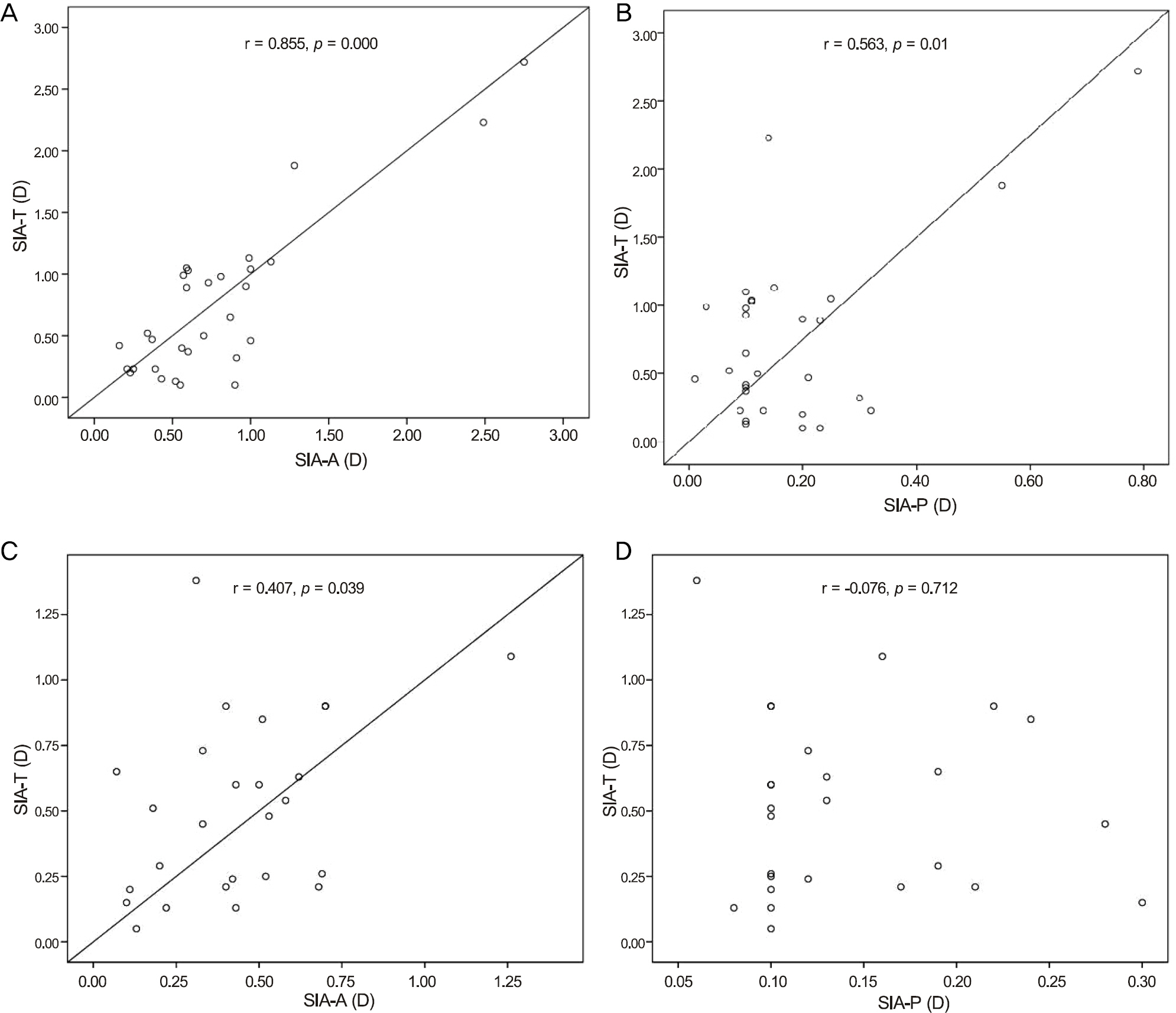
Figure 2. Correlation of magnitude of SIA-A, SIA-P and SIA-T in CCS group (A,B) and MCCS group (C,D). SIA-A = surgically induced astigmatism-anterior; SIA-P = surgically induced astigmatism-posterior; SIA-T = surgically induced astigmatism-total; CCS = conventional cataract surgery; MCCS = microcoaxial cataract surgery.
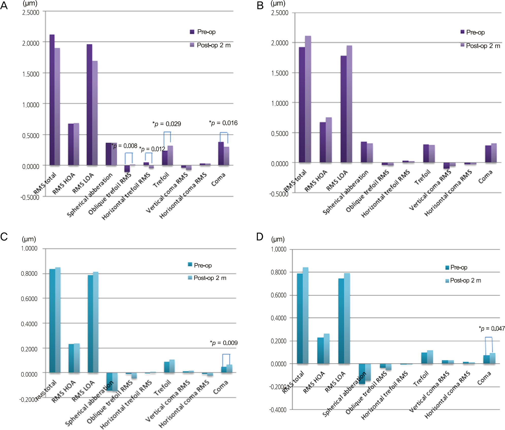
Figure 3. Change in corneal higher order aberrations on front side in CCS group (A) and MCCS group (B) and on rear side in CCS group (C) and MCCS group (D). CCS = conventional cataract surgery; MCCS = microcoaxial cataract surgery; RMS = root mean square; HOA = high order abberation; LOA = lower order aberrations; Pre-op = preoperative; Post-op 2 m = postoperative 2 months. * p-value by Paired t-test ( p < 0.05).
Reference
-
References
1. Wolffsohn JS, Bhogal G, Shah S. Effect of uncorrected astigma-tism on vision. J Cataract Refract Surg. 2011; 37:454–60.
Article2. de Vries NE, Webers CA, Touwslager WR. . Dissatisfaction af-ter implantation of multifocal intraocular lenses. J Cataract Refract Surg. 2011; 37:859–65.
Article3. Amesbury EC, Miller KM. Correction of astigmatism at the time of cataract surgery. Curr Opin Ophthalmol. 2009; 20:19–24.
Article4. Altan-Yaycioglu R, Akova YA, Akca S. . Effect on astigma-tism of the location of clear corneal incision in phacoemulsifica-tion of cataract. J Refract Surg. 2007; 23:515–8.
Article5. Hill W. Expected effects of surgically induced astigmatism on AcrySof toric intraocular lens results. J Cataract Refract Surg. 2008; 34:364–7.
Article6. Borasio E, Mehta JS, Maurino V. Surgically induced astigmatism after phacoemulsification in eyes with mild to moderate corneal as-tigmatism: temporal versus on-axis clear corneal incisions. J Cataract Refract Surg. 2006; 32:565–72.7. Dick HB. Controlled clinical trial comparing biaxial microincision with coaxial small incision for cataract surgery. Eur J Ophthalmol. 2012; 22:739–50.
Article8. Alió J, Rodríguez-Prats JL, Galal A, Ramzy M. Outcomes of mi-croincision cataract surgery versus coaxial phacoemulsification. Ophthalmology. 2005; 112:1997–2003.
Article9. Koch DD, Ali SF, Weikert MP. . Contribution of posterior cor-neal astigmatism to total corneal astigmatism. J Cataract Refract Surg. 2012; 38:2080–7.
Article10. Cheng LS, Tsai CY, Tsai RJ. . Estimation accuracy of surgically induced astigmatism on the cornea when neglecting the posterior corneal surface measurement. Acta Ophthalmol. 2011; 89:417–22.
Article11. Ho JD, Tsai CY, Liou SW. Accuracy of corneal astigmatism esti-mation by neglecting the posterior corneal surface measurement. Am J Ophthalmol. 2009; 147:788–95. 795.e1-2.
Article12. Bhargava S, Rangarajan A. Coaxial microincision cataract surgery versus conventional coaxial cataract surgery. J Cataract Refract Surg. 2008; 34:1055. author reply 1055.
Article13. Weikert MP. Update on bimanual microincisional cataract surgery. Curr Opin Ophthalmol. 2006; 17:62–7.
Article14. Nemeth G, Berta A, Lipecz A. . Evaluation of posterior astig-matism measured with Scheimpflug imaging. Cornea. 2014; 33:1214–8.
Article15. Elkady B, Alió JL, Ortiz D, Montalbán R. Corneal aberrations after microincision cataract surgery. J Cataract Refract Surg. 2008; 34:40–5.
Article16. Tong N, He JC, Lu F. . Changes in corneal wavefront aberra-tions in microincision and small-incision cataract surgery. J Cataract Refract Surg. 2008; 34:2085–90.
Article17. Yao K, Tang X, Ye P. Corneal astigmatism, high order aberrations, and optical quality after cataract surgery: microincision versus small incision. J Refract Surg. 2006; 22:(9 Suppl). S1079–82.
Article18. Guirao A, Tejedor J, Artal P. Corneal aberrations before and after small-incision cataract surgery. Invest Ophthalmol Vis Sci. 2004; 45:4312–9.
Article19. Marcos S, Rosales P, Llorente L, Jiménez-Alfaro I. Change in cor-neal aberrations after cataract surgery with 2 types of aspherical in-traocular lenses. J Cataract Refract Surg. 2007; 33:217–26.
Article20. Wang J, Tang X, Zhang S, Li LH. Changes in high order aberra-tions of anterior and posterior surfaces of cornea before and after phacoemulsification. Zhonghua Yan Ke Za Zhi. 2008; 44:1066–71.
- Full Text Links
-
- Actions
-
Cited
- CITED
-
- Close
- Share
-
- Similar articles
-
- Comparative Study of Microcoaxial Cataract Surgery and Conventional Cataract Surgery
- Induced Astigmatism and High-Order Aberrations after 1.8-mm, 2.2-mm and 3.0-mm Coaxial Phacoemulsification Incisions
- One-Year Outcome of Microcoaxial Cataract Surgery Using 1.8 mm and 2.2 mm Incisions Versus that of Conventional Cataract Surgery
- Surgically Induced Astigmatism and Corneal Higher Order Aberrations in Microcoaxial and Conventional Cataract Surgery
- Comparison of Phacodynamic Effects on Postoperative Corneal Edema Between 2.8 mm and 2.2 mm Microcoaxial Torsional Phacoemulsification
