Wavefront Analysis of Successful Treatment of Monocular Triplopia After Cataract Extraction: Report of 2 Cases
- Affiliations
-
- 1Department of Ophthalmology, Ilsan Paik Hospital, Inje University College of Medicine, Goyang, Korea. eyedr0823@hotmail.com
- KMID: 2213791
- DOI: http://doi.org/10.3341/jkos.2010.51.7.1003
Abstract
- PURPOSE
To report wavefront analysis of successful treatment of monocular triplopia after cataract extraction.
CASE SUMMARY
(Case 1) A 59-year-old man visited our clinic for a monocular triplopia in his left eye of five years in duration. The best spectacle-corrected visual acuity (BSCVA) was 1.0 in the left eye, and the patient had a mild cortical cataract. The ocular spherical aberration (0.126 micrometer for the 4-mm pupil, 0.351 micrometer for the 6-mm pupil) measured by a Hartmann-Shack aberrometer increased preoperatively, while the corneal spherical aberration was normal. After cataract surgery, the monocular triplopia disappeared, and the ocular spherical aberration decreased. (Case 2) A 38-year-old man visited our clinic for a monocular triplopia in his left eye of a two-year duration. The best spectacle-corrected visual acuity (BSCVA) was 0.3 in the left eye, and the patient had a mild nuclear cataract. The ocular spherical aberration (-0.356 micrometer, -1.343 micrometer) and trefoil aberration (0.199 micrometer, 0.252 micrometer) increased preoperatively, while the corneal spherical and trefoil aberrations were normal. After cataract surgery, the monocular triplopia disappeared and the ocular spherical and trefoil aberrations decreased.
Keyword
Figure
-
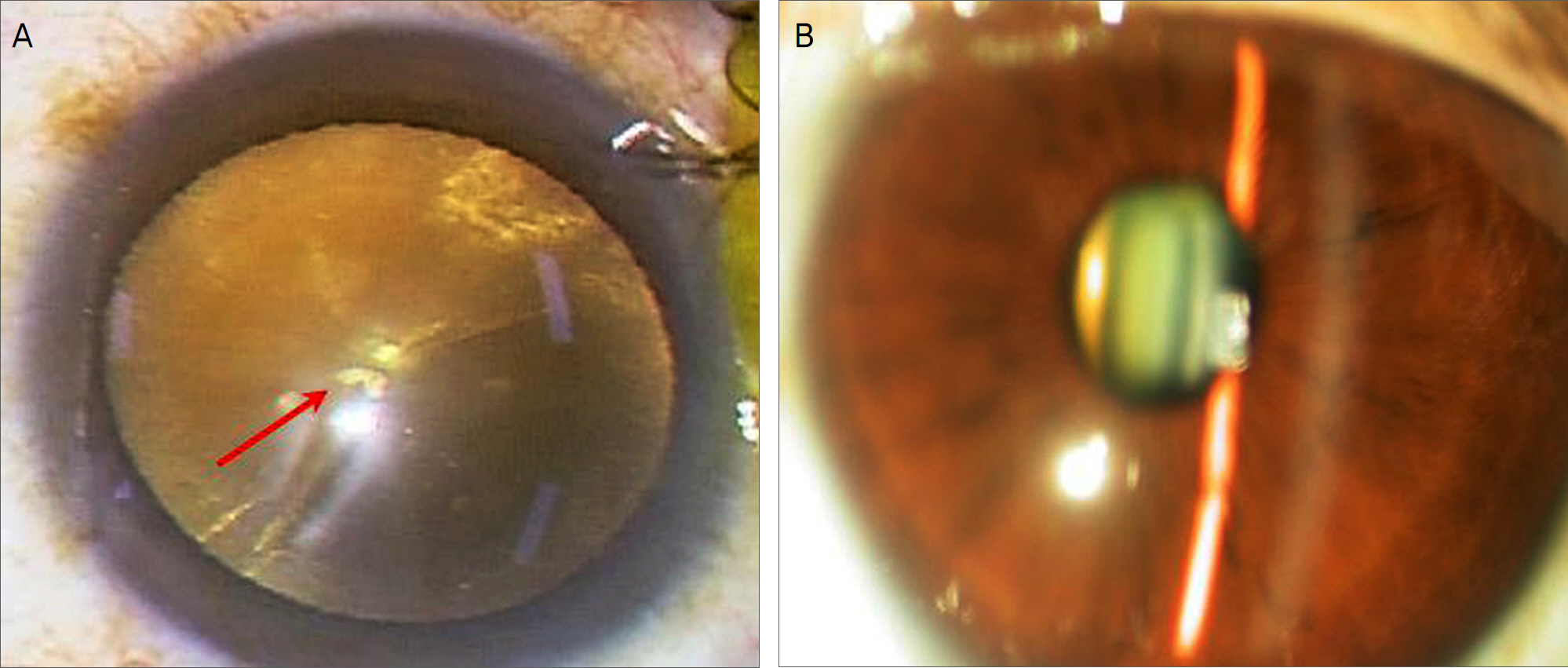
Figure 1. Preoperative photograph. (Case 1) Intraoperative photograph reveals some cystic vacuoles involving the center of the pupil (red arrow) and mild cortical cataract (A). (Case 2) Slit lamp photograph shows mild nuclear cataract (B).
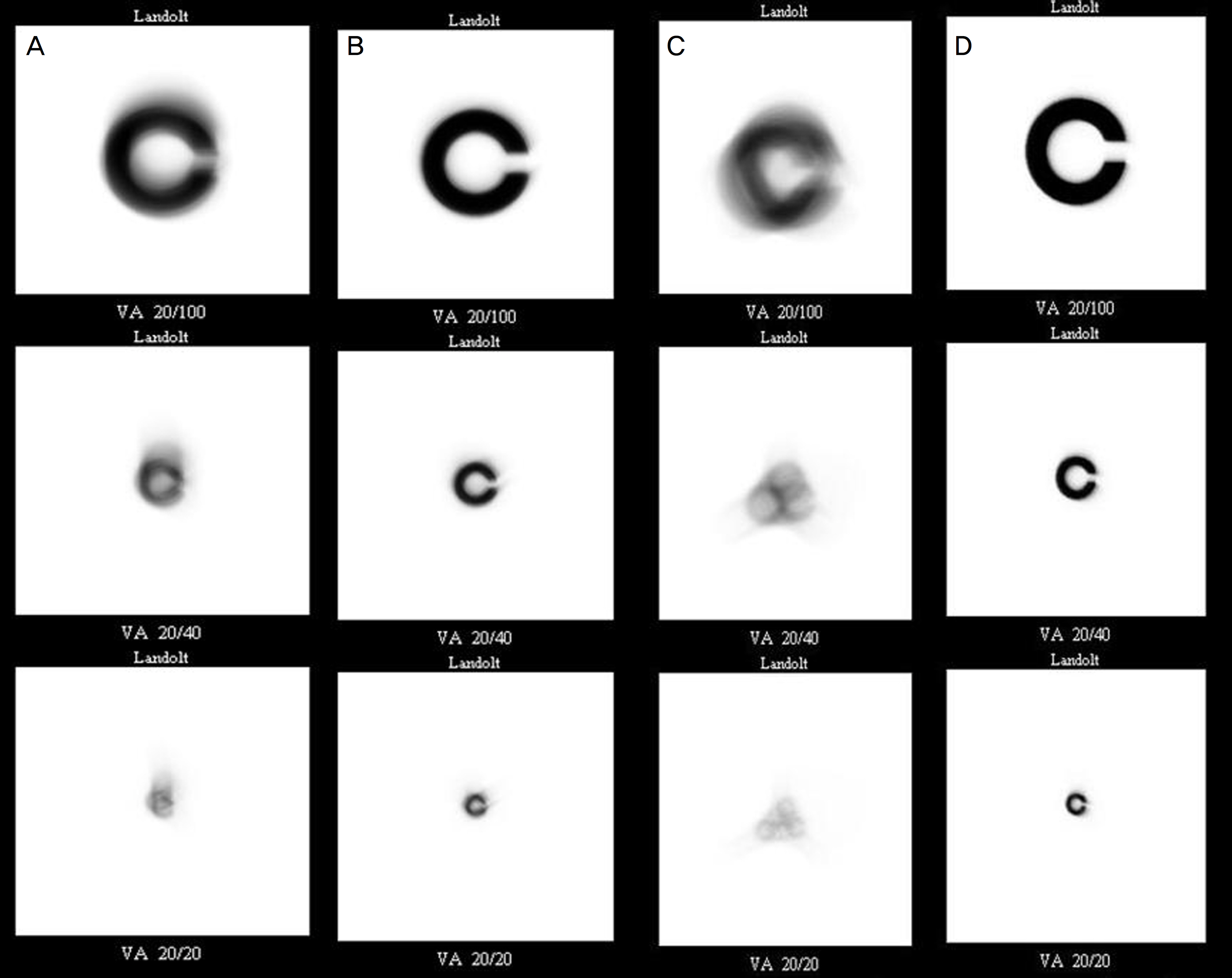
Figure 2. Simulated image for Landolt C before and after surgery. (Case1) Simulated image for Landolt C shows triple configuration before surgery (A). However, simulated image for Landolt C shows normal pattern after surgery (B). (Case2) Simulated image for Landolt C shows triple configuration before surgery (C). However, simulated image for Landolt C shows normal pattern after surgery (D).
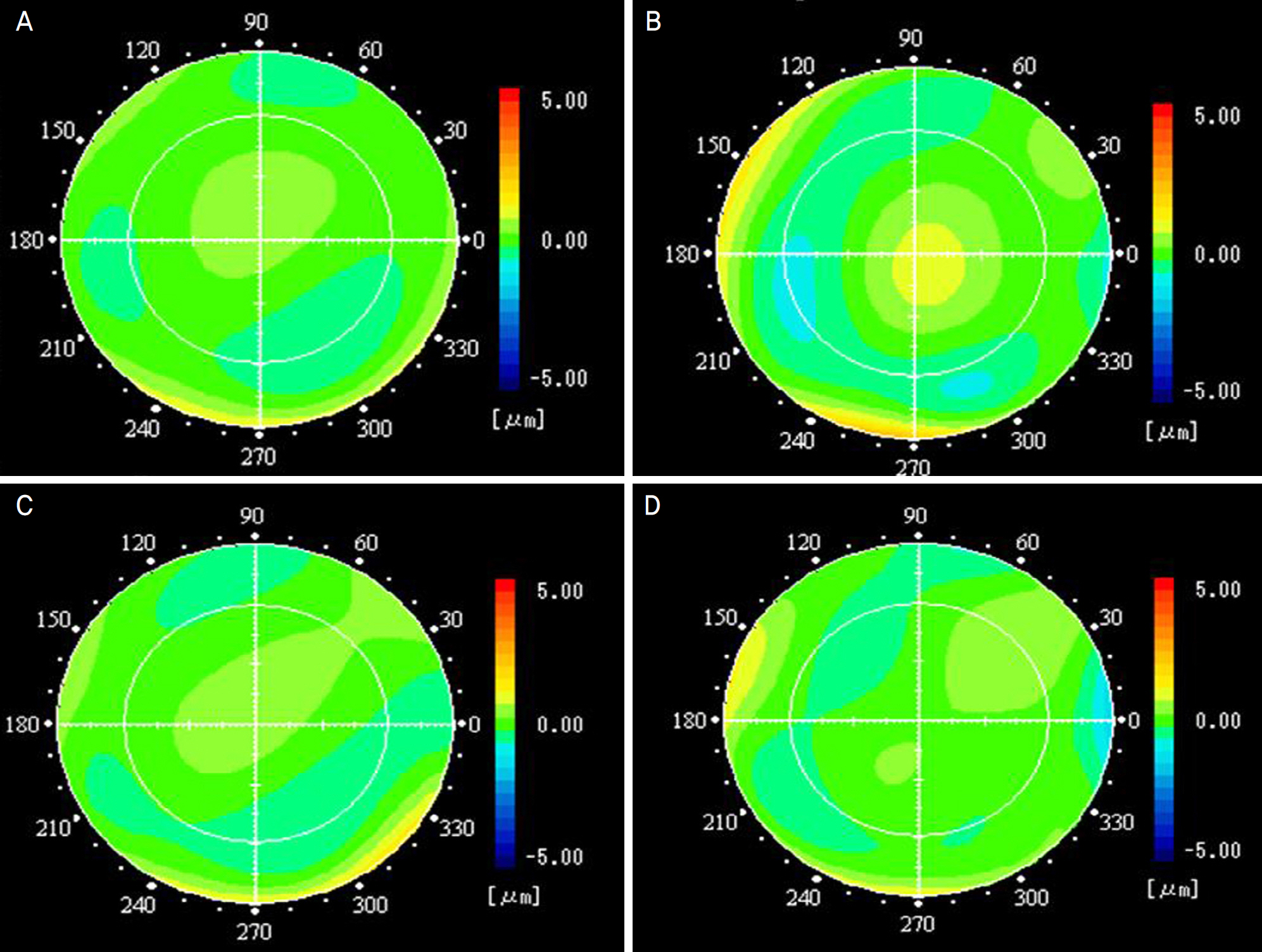
Figure 3. (Case1) Wavefront analysis of higher-order aberration in the cornea and in oculus before and after surgery. Corneal higher-order aberrations had almost normal pattern before and after surgery (A, C), but ocular higher-order aberrations showed advancement of wavefront (warm color) in the central pupillary area and delay of wavefront (cool color) in peripheral pupillary area before surgery (B) and a normal pattern after surgery (D).
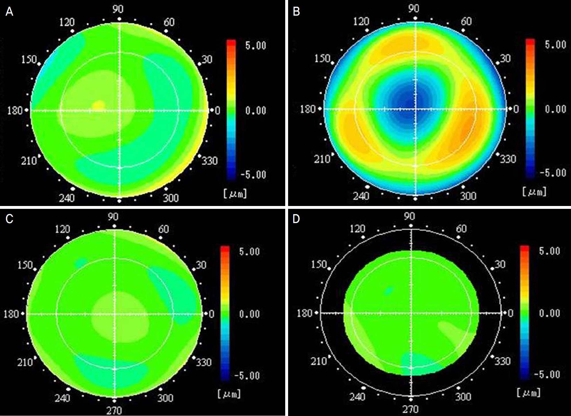
Figure 4. (Case 2) Wavefront analysis of higher-order aberration in the cornea and in oculus before and after surgery. Corneal higher-order aberrations had almost normal pattern before and after surgery (A, C), but ocular higher-order aberrations showed a delay of wavefront (cool color) in the central pupillary area and trefoil pattern in the peripheral area before surgery (B) and a normal pattern after surgery (D).
Reference
-
References
1. Coffeen P, Guyton DL. Monocular diplopia accompanying ordinary refractive errors. Am J Ophthalmol. 1998; 105:451–9.
Article2. Bowman KJ, Smith G, Carney LG. Corneal topography and monocular diplopia following near work. Am J Optom Physiol Opt. 1978; 55:818–23.
Article3. Fincham EF. Monocular diplopia. Br J Ophthalmol. 1963; 47:705–12.
Article4. Kuroda T, Fujikado T, Maeda N, et al. Wavefront analysis in eyes with nuclear or cortical cataract. Am J Ophthalmol. 2002; 134:1–9.
Article5. Fujikado T, Shimojyo H, Hosohata J, et al. Wavefront analysis of eye with monocular diplopia and cortical cataract. Am J Ophthalmol. 2006; 141:1138–40.
Article6. Kim A, Bessho K, Okawa Y, et al. Wavefront analysis of eyes with cataracts in patients with monocular triplopia. Ophthalmic Physiol Opt. 2006; 26:65–70.7. Kuroda T, Fujikado T, Maeda N, et al. Wavefront analysis of high-er-order aberrations in patients with cataract. J Cataract Refract Surg. 2002; 28:438–44.
Article8. Fujikado T, Kuroda T, Maeda N, et al. Wavefront analysis of an eye with monocular triplopia and nuclear cataract. Am J Ophthalmol. 2004; 137:361–3.
Article9. Yoon G, Jeong TM, Cox IG, Williams DR. Vision improvement by correcting higher-order aberrations with phase plates in normal eyes. J Refract Surg. 2004; 20:523–7.
Article10. Oie Y, Maeda N, Kosaki R, et al. Characteristics of ocular high-er-order aberrations in patients with pellucid marginal corneal degeneration. J Cataract Refract Surg. 2008; 34:1928–34.
Article
- Full Text Links
-
- Actions
-
Cited
- CITED
-
- Close
- Share
-
- Similar articles
-
- The Measurement of the Laser Space in the Planned Extracapsular Cataract Extraction with the PCL
- The Complications of Cataract Extraction
- Clinical Studies on Hyphema in The Complications of Cataract Extraction
- Change in Angle Deviation After Visual Acuity Improvement in Monocular Deviated Patients
- The Change of the Curneal Curvature Before and After Cataract Operation
