J Korean Ophthalmol Soc.
2009 Oct;50(10):1510-1513. 10.3341/jkos.2009.50.10.1510.
Clinical Features of Conjunctival Nevi in Korean Patients
- Affiliations
-
- 1Kim's Eye Hospital, Seoul, Korea.
- 2Department of Ophthalmology, Seoul National University College of Medicine Seoul Artificial Eye Center, Clinical Research Institute, Seoul National University Hospital, Seoul, Korea. eyeminerva@yahoo.co.kr
- 3Seoul National University Hospital Health Care System Gangnam Center, Healthcare Reaserch Institute, Seoul, Korea.
- KMID: 2212768
- DOI: http://doi.org/10.3341/jkos.2009.50.10.1510
Abstract
- PURPOSE
To evaluate clinical features and therapeutic modality of conjunctival nevi in Korean patients.
METHODS
A retrospective analysis was performed on 197 patients (75 males and 122 females) with nevi who were diagnosed by slit lamp examination from 1997 to 2008.
RESULTS
Nevi occurred most commonly on bulbar conjunctiva (88%), followed by caruncle and plica semilunaris (7%). The nevi involved temporal (71%), nasal (21%), inferior (2.8%) and superior (0.7%) quadrants of the conjunctiva. The mean horizontal length was 4.3+/-2.0 mm and the mean vertical 4.45+/-2.2 mm. Thirty-five patients (7.8%) received no treatment. Excisional biopsy was performed in 38 patients (19.3%). Argon laser photoablation of conjunctiva nevi was performed in 124 patients (62.9%).
CONCLUSIONS
The pattern of conjunctival nevi in Korean patients was similar to Caucasian patients. The biopsy was performed according to appropriate guidelines however, nevus which does not require a biopsy, could be treated by argon laser photoablation.
Figure
-
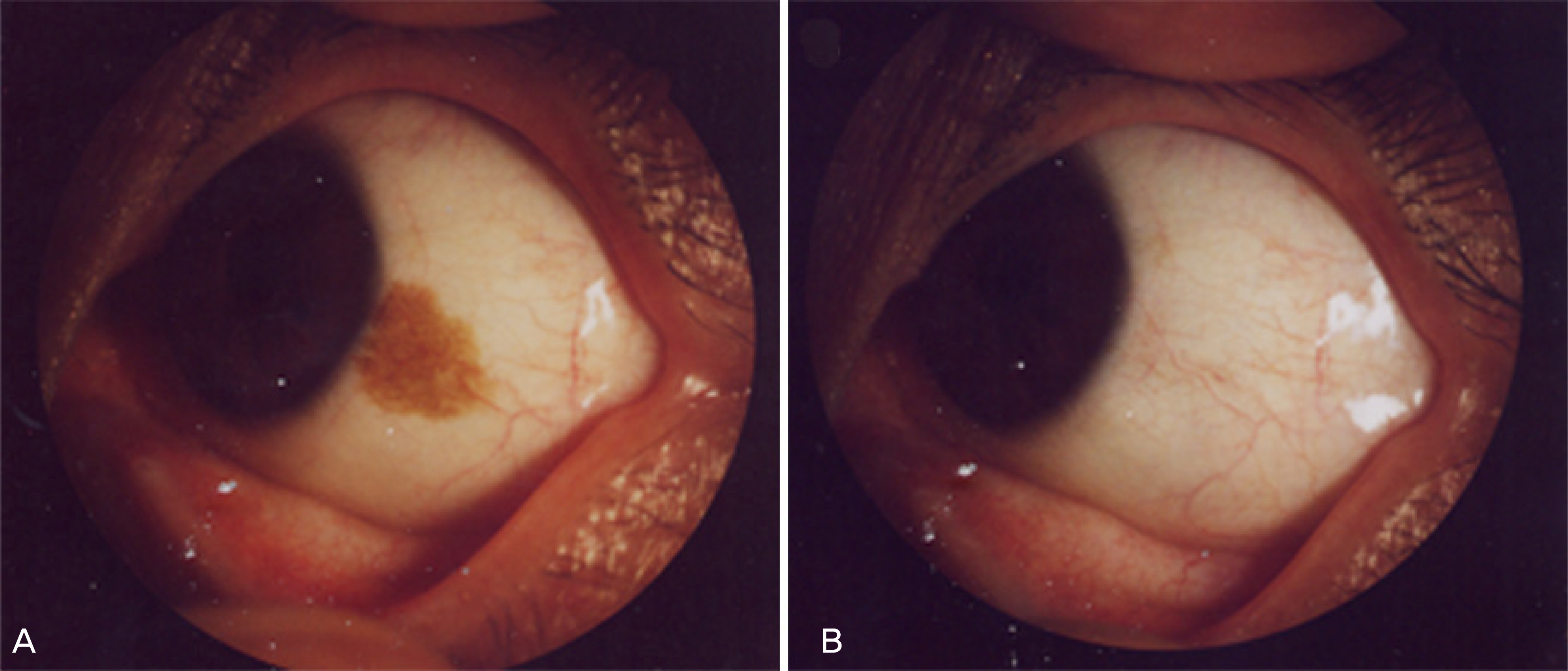
Figure 1. Conjunctival nevus before argon laser photoablation (A) and after laser treatment (B).
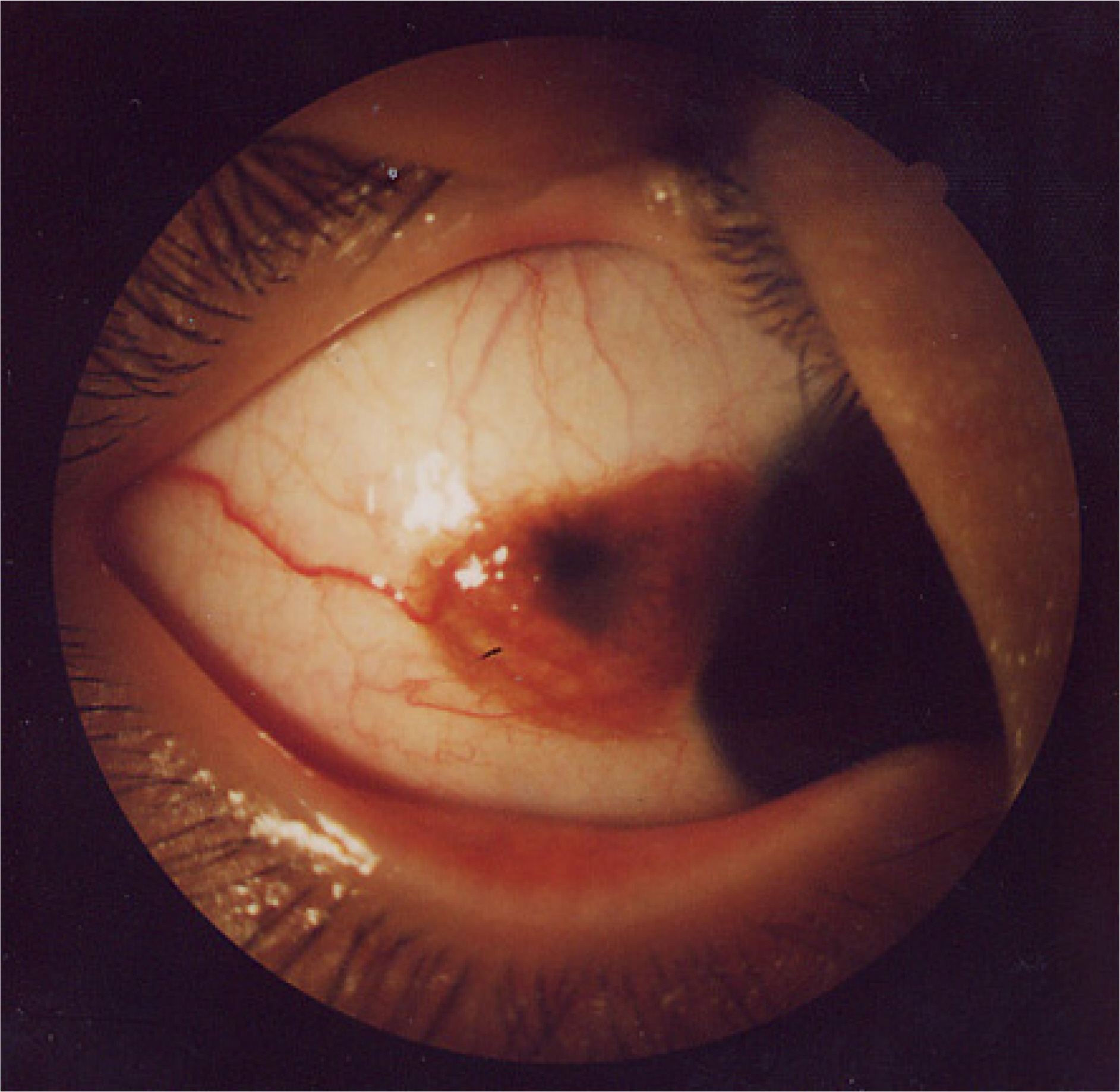
Figure 2. Compound nevus with a feeder vessel.
Cited by 1 articles
-
Surgical Outcome of Chemical Peeling of Conjunctival Nevus with Alcohol
Wong Bong Jang, Sang Jun Ko, Sang Duck Kim
J Korean Ophthalmol Soc. 2016;57(5):705-709. doi: 10.3341/jkos.2016.57.5.705.
Reference
-
References
1. Amoli FA, Heidari AB. Survey of 447 patient with conjunctival neoplastic lesions in Farabi eye hospital, Teheran, Iran. Ophthalmic Epidemiol. 2006; 13:275–9.2. Shields CL, Fasiudden A, Mashayekhi A, Shields JA. Conjuntival nevi: Clinical Features and Natural Course in 410 Consecutive Patients. Arch Ophthalmol. 2004; 122:167–75.3. Kurli M, Finger PT. Melanocytic conjunctival tumors. Ophthalmol Clin North Am. 2005; 18:15–24.
Article4. Thiagalingam S, Johnson MM, Colby KA, Zembowicz A. Juvenile conjunctival nevus. Am J Surg Pathol. 2008; 32:399–406.
Article5. Jeoung JW, Kim T, Lee JH, et al. Argon laser ablation of conjunctival nevus. J Korean Ophthalmol Soc. 2004; 45:1989–94.
