J Korean Ophthalmol Soc.
2009 Jan;50(1):14-18. 10.3341/jkos.2009.50.1.14.
Success Rates According to the Shape of Rhinostomy After Endonasal Dacryocystorhinostomy
- Affiliations
-
- 1Myung Gok Eye Research Institute, Kim's Eye Hospital, Konyang University, Seoul, Korea. jjw@kimeye.com
- 2Department of Ophthalmology, Dongguk University College of Medicine, Gyeongju, Korea.
- 3Department of Ophthalmology, College of Medicine, Konyang University, Daejeon, Korea.
- KMID: 2211892
- DOI: http://doi.org/10.3341/jkos.2009.50.1.14
Abstract
- PURPOSE
To evaluate the success rates according to the shape of rhinostomy after endonasal dacryocystorhinostomy.
METHODS
We analyzed 131 patients (188 cases) who had undergone endonasal dacryocystorhinostomy with a minimum follow-up of 6 months. After surgery, all cases were classified into flat, alcove, concave, or concealed concave types according to Jane's rhinostomy classification. Patency to irrigation and improved symptoms were considered indicative of a successful outcome.
RESULTS
There were 81 flat type patients (125 cases), 31 alcove type patients (42 cases), 19 concave type patients (21 cases), and no cases were classified as concealed concave type. The success rates were 75.2%, 85.7%, 81.0% respectively, and the most common cause of failure was the formation of granulation within the ostium. The success rates did not significantly vary according to the shape of rhinostomy (p=0.144).
CONCLUSIONS
The success rates after endonasal dacryocystorhinostomy of flat, alcove, and concave types in turn were 75.2%, 85.7%, and 81.0%.
MeSH Terms
Figure
-
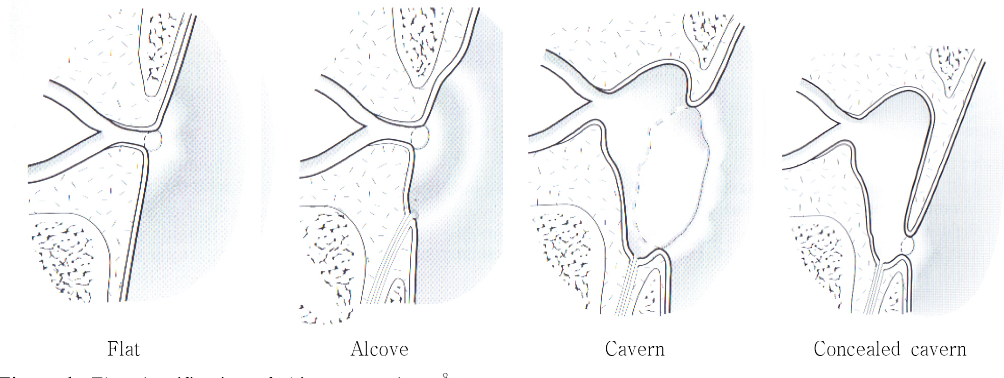
Figure 1. The classification of rhinostomy shape.8

Figure 2. Transnasal endoscopic photographs of flat (A), alcove (B), and cavern (C) shape at postoperative 3 months.
Cited by 2 articles
-
Comparison of Drills Used in Endonasal Dacryocystorhinostomy
Sang Won Moon, Yoon Jung Lee
J Korean Ophthalmol Soc. 2010;51(4):479-484. doi: 10.3341/jkos.2010.51.4.479.Surgical Outcomes of Endonasal Dacryocystorhinostomy According to the Level of Obstruction in Dacryocystography
Kyoung Hwa Bae, Nam Chun Cho, Min Ahn
J Korean Ophthalmol Soc. 2018;59(4):301-306. doi: 10.3341/jkos.2018.59.4.301.
Reference
-
References
1. Goldberg RA. Endonasal Dacryocystorhinostomy: is it really less successful? Arch Opthalmol. 2004; 122:108–10.2. Park JD, Kim YI, Shin SG. The factors related to surgical success rate of endonasal dacryocystorhinostomy. J Korean Ophthalmol Soc. 1998; 39:10–5.3. Lee DP, Yang SW, Choi WC. The relation between nasal cavity size and success rate in endonasal dacryocystorhino-stomy. J Korean Ophthalmol Soc. 2000; 41:86–91.4. Mannor GE, Millman AL. The prognostic value of pre-operative dacryocystography in endoscopic intranasal dacryo-cystorhinostomy. Am J Ophthalmol. 1992; 113:134–7.
Article5. Lee HC, Chung WS. Success rate of endonasal dacryocy-storhinostomy. J Korean Ophthalmol Soc. 1996; 37:211–8.6. Fayet B, Racy E, Assouline M. Complications of Standardized Endonasal Dacryocystorhinostomy with Unciformectomy. Ophthalmology. 2004; 111:837–45.7. Metson R. Endoscopic surgery for lacrimal obstruction. Oto-laryngol Head Neck Surg. 1991; 104:473–9.
Article8. Jane Olver. Colour Atlas of Lacrimal Surgery. Oxford: Butter-worth Heinemann;2002. p. 135.9. Chung ER, Lee KT, Choi WC. Success rate of endonasal dacryocystorhinostomy based on the location of the lacrimal sac. J Korean Ophthalmol Soc. 2002; 43:2000–4.10. Kang IB, Kim ST, Kim CW. . Endoscopic laser dacryo-cystorhinostomy. J Korean Otolaryngol Soc. 1998; 41:746–9.11. Bumstead RM, Linberg JV, Anderson RL, Barreras R. External Dacryocystorhinostomy. A Prospective Study Comparing the Size of the Operative and Healed Ostium. Arch Otolaryngol. 1982; 108:407–10.12. Lee TS, Kim SW, Park BW. The Relationship between Rate of Wound Healing and Success Rate after Endonasal Laser-drill Assisted Dacryocystorhinostomy. J Korean Ophthalmol Soc. 1999; 40:2969–74.13. Jin HR, Shin SO, Choi YS. . Endoscopic Endonasal Dacryocystorhinostomy: Prevention of Neo-Ostium Obstruction Using Nasal Mucosal Flap. J Korean Otolaryngol Soc. 2003; 46:1040–5.14. Choi YJ, Hwang SJ, Lee TS. Short-Term Clinical Results of Amniotic Membrane Application to Endonasal Dacryocy-storhinostomy. J Korean Ophthalmol Soc. 2008; 29:384–9.
Article15. Park DJ, Kwak MS. The Effect of Mitomycin C on Success Rate of Endoscopic Dacryocystorhinostomy. J Korean Ophthalmol Soc. 2000; 41:1674–9.16. Woog JJ, Kennedy RH, Custer PL. . Endonasal Dacryo-cystorhinostomy. Ophthalmology. 2001; 108:2369–77.
Article17. Linberg JV, Anderson RL, Bumsted RM, Barreras R. Study of Intranasal Ostium External Dacryocystorhinostomy. Arch Ophthalmol 100. 1982:1758–62.
Article18. Fayet B, Racy E, Assouline M, Zerbib M. Surgical Anatomy of the Lacrimal Fossa: A Prospective Computed Tomodensito-metry Scan Analysis. Ophthalmology. 2005; 112:1119–28.19. Yazici B, Yazici Z. Anatomic position of the common canaliculus in patients with a large lacrimal sac. Ophthalm Plast Reconstr Surg. 2008; 24:90–3.20. Lee KC, Kwon KW. Posterior Sac Approach in Endoscopic Dacryocystorhinostomy. J Korean Otolaryngol Soc. 2000; 43:213–6.21. Fayet B, Racy E, Assouline M. Systematic Uniformectomy for a Standardized Endonasal Dacryocystorhinostomy. Ophthalmology. 2002; 109:530–36.22. Lee SG, Yang JW, Shin SG, Choi MG. Effect of Lacrimal Sac Incision Using Keratome in Endonasal Dacryocystorhinostomy. J Korean Ophthalmol Soc. 2003; 44:2720–6.23. Oh IK, Cho SH, Lee TS. Comparison of Results between.
- Full Text Links
-
- Actions
-
Cited
- CITED
-
- Close
- Share
-
- Similar articles
-
- Clinical Evaluation of Endoscopic Endonasal Dacryocystorhinostomy
- Success Rate of Endonasal Dacryocystorhinostomy Based on the Location of the Lacrimal Sac
- The Effect of Nasal Cavity Abnormality Related to Surgical Success Rate of Endonasal Dacryocystorhinostomy
- Clinical Experience of Lacrimal Surgery
- Silicone Tube Intubation with Lacrimal Endoscopy and Endonasal Dacryocystorhinostomy in Adult Nasolacrimal Duct Obstruction
