J Korean Ophthalmol Soc.
2008 May;49(5):798-810. 10.3341/jkos.2008.49.5.798.
Diagnostic Ability of Stratus OCT Using Korean Normative Database for Early Detection of Normal-Tension Glaucoma
- Affiliations
-
- 1Department of Ophthalmology, Hanyang University College of Medicine, Seoul, Korea. KBUhm@hanyang.ac.kr
- 2Lee's Eye Clinic, Hongsung, Korea.
- KMID: 2211711
- DOI: http://doi.org/10.3341/jkos.2008.49.5.798
Abstract
-
PURPOSE: To determine if korean normative database improve diagnostic ability of Stratus OCT for detection of glaucoma.
METHODS
Peripapillary retinal nerve fiber layer (RNFL) and optic nerve head regions were measured using a Stratus OCT. The normative data were collected from 129 normal individuals. We obtained values at the 5% and 1% levels according to the disc area stratified into equal thirds to minimize the error by the individual variation of optic disc size and these levels were considered abnormal. One eye of each 94 normal-tension glaucoma patients with early visual field defects (mean deviaton = -3.84+/-1.40dB) and 87 another normal subjects were enrolled. Glaucoma was defined by visual field defects.
RESULTS
The use of Korean normative database had higher sensitivity and no significant difference of specificity than that of a Stratus OCT except for a few parameters. The criteria that show the highest diagnostic ability were 1 > or = quadrants RNFL thickness abnormal at the < 5% when using a Korean normative database (sensitivity = 81.9%, specificity = 81.6%) and 1 > or = clock hours abnormal at the < 5% when using a Stratus OCT normative database (sensitivity = 71.3%, specificity = 87.4%) (p=0.004, p=0.180, respectively, McNemar test).
CONCLUSIONS
The criterion using a Korean normative database had higher sensitivity and no difference of specificity compared with criterion using a Stratus OCT normative database. The best criterion using a Korean normative database may be helpful for early detection of normal-tension glaucoma.
MeSH Terms
Figure
-
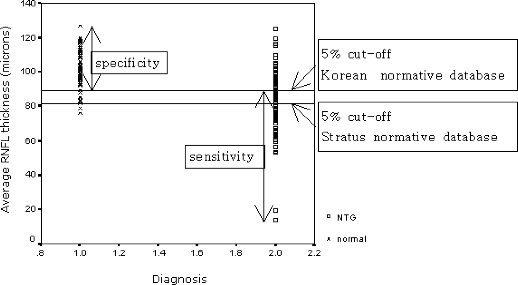
Figure 1. The scatter diagram of average retinal nerve fiber layer thickness (RNFL) in 87 normal subjects and 94 early normal tension glaucoma (NTG) patients. The use of 5% cut off values from a Korean normative database had higher sensitivity than that from a Stratus OCT.
Cited by 1 articles
-
False Negative Findings of Optical Coherence Tomography in Eyes with Localized Nerve Fiber Layer Defects
Sung Min Kang, Ki Bang Uhm
J Korean Ophthalmol Soc. 2011;52(4):454-461. doi: 10.3341/jkos.2011.52.4.454.
Reference
-
References
1. Quigley HA, Addicks EM, Green WR. Optic nerve damage in human glaucoma. III. Quantitative correlation of nerve fiber loss and visual field defect in glaucoma, ischemic neuropathy, papilledema, and toxic neuropathy. Arch Ophthalmol. 1982; 100:135–46.2. Javitt JC, Spaeth CL, Katz J, et al. Acquired pits of the optic nerve. Increased prevalence in patients with low tension glaucoma. Ophthalmology. 1990; 97:1038–44.3. Broadway DC, Grierson I, Hitchings RA. The ability of scanning laser ophthalmoscopy to identify various glaucomatous optic disk appearances. Am J Ophthalmol. 1998; 125:593–604.
Article4. Quigley HA, Brown AE, Morrison JD, Drance SM. The size and shape of the optic disc in normal human eyes. Arch Ophthalmol. 1990; 108:51–7.
Article5. Poinoosawmy D, Fontana L, Wu JX, et al. Variation of nerve fiber layer thickness measurements with age and ethnicity by scanning laser polarimetry. Br J Ophthalmol. 1997; 81:350–4.6. Iester M, Mikelberg FS, Drance SM. The effect of optic disc size on diagnostic precision with the Heidelberg retina tomograph. Ophthalmology. 1997; 104:545–8.
Article7. Savini G, Zanini M, Carelli V, et al. Correlation between retinal nerve fiber layer thickness and optic nerve head size: an optical coherence tomography study. Br J Ophthalmol. 2005; 89:489–92.8. Bowd C, Zangwill LM, Blumenthal EZ, et al. Imaging of the optic disc and retinal nerve fiber layer: the effects of age, optic disc area, refractive error, and gender. J Opt Soc Am A Opt Image Sci Vis. 2002; 19:197–207.
Article9. Jonas JB, Schmidt AM, Muller-Bergh JA, et al. Human optic nerve fiber count and optic disc size. Invest Ophthalmol Vis Sci. 1992; 33:2012–8.10. Garway-Heath DF, Hitchings RA. Sources of bias in studies of optic disc and retinal nerve fiber layer morphology. Br J Ophthalmol. 1998; 82:986.11. Paunescu LA, Schuman JS, Price LL, et al. Reproducibility of nerve fiber thickness, macular thickness, and optic nerve head measurements using Stratus OCT. Invest Ophthalmol Vis Sci. 2004; 45:1716–24.12. Medeiros FA, Zangwill LM, Bowd C, et al. Influence of disease severity and optic disc size on the diagnostic performance of imaging instruments in glaucoma. Invest Ophthalmol Vis Sci. 2006; 47:1008–15.
Article13. Uhm KB, Lee JS. Optic disc measurements with personal computer in normal eyes. J Korean Ophthalmol Soc. 1995; 36:1760–9.14. Park HJ, Hwang JH, Uhm KB. The influence of age, gender, refractive error, and optic disc area on the HRT parameters in normal eyes. J Korean Ophthalmol Soc. 2004; 45:982–9.15. Han JI, Lim HW, Song YM, Uhm KB. Factors influencing optic disc and retinal nerve fiber layer parameters measured by optical coherence tomography. J Korean Ophthalmol Soc. 2007; 48:1073–81.
Article16. Budenz DL, Anderson DR, Varma R, et al. Determinants of normal retinal nerve fiber layer thickness measured by Stratus OCT. Ophthalmology. 2007; 114:1046–52.
Article17. Brusini P, Salvetat ML, Zeppieri M, et al. Comparison between GDx VCC scanning laser polarimetry and Stratus OCT optical coherence tomography in the diagnosis of chronic glaucoma. Acta Ophthalmol Scand. 2006; 84:650–5.
Article18. Chen HY, Huang ML. Discrimination between normal and glaucomatous eyes using Stratus optical coherence tomography in Taiwan Chinese subjects. Graefes Arch Clin Exp Ophthalmol. 2005; 243:894–902.
Article19. Medeiros FA, Zangwill LM, Bowd C, et al. Evaluation of retinal nerve fiber layer, optic nerve head, and macular thickness measurements for glaucoma detection using optical coherence tomography. Am J Ophthalmol. 2005; 139:44–55.
Article20. Wollstein G, Ishikawa H, Wang J, et al. Comparison of three optical coherence tomography scanning areas for detection of glaucomatous damage. Am J Ophthalmol. 2005; 139:39–43.
Article21. Manassakorn A, Nouri-Mahdavi K, Caprioli J. Comparison of retinal nerve fiber layer thickness and optic disk algorithms with optical coherence tomography to detect glaucoma. Am J Ophthalmol. 2006; 141:105–15.
Article22. Kang KD, Park CK. Comparison of diagnostic precision between preprogramed indicator and newly calculated indicator in optical coherence tomography. J Korean Ophthalmol Soc. 2006; 47:243–52.23. Budenz DL, Michael A, Chang RT, et al. Sensitivity and specificity of the Stratus OCT for perimetric glaucoma. Ophthalmology. 2005; 112:3–9.24. Kim JM, Park KH, Kim TW, Kim DM. Comparison of the results between Heidelberg Retina Tomography II and Stratus optical coherence tomography in glaucoma. J Korean Ophthalmol Soc. 2006; 47:556–62.25. Hougaard JL, Kessel L, Sander B, et al. Evaluation of heredity as a determinant of retinal nerve fiber layer thickness as measured by optical coherence tomography. Invest Ophthalmol Vis Sci. 2003; 44:3011–6.
Article
- Full Text Links
-
- Actions
-
Cited
- CITED
-
- Close
- Share
-
- Similar articles
-
- Comparison of Diagnostic Ability of 3D and Stratus Optical Coherence Tomography in Early Glaucoma
- Comparison of the Results between Heidelberg Retina Tomography II and Stratus Optical Coherence Tomography in Glaucoma
- Comparison of RNFL Thickness Measured by Two Different Kind of OCT in NTG Patients
- Discrimination between Normal and Early Stage of Glaucomatous Eyes Using the Stratus Optical Coherence Tomography
- Stratus OCT, SWAP, Matrix FDT in Preperimetric Glaucoma
