J Korean Surg Soc.
2009 Feb;76(2):127-130. 10.4174/jkss.2009.76.2.127.
Laparoscopic Treatment of Mesenteric Castleman's Disease
- Affiliations
-
- 1Department of Surgery, Daejeon St. Mary's Hospital, The Catholic University of Korea College of Medicine, Daejeon, Korea. leekj@catholic.ac.kr
- 2Department of Pathology, Daejeon St. Mary's Hospital, The Catholic University of Korea College of Medicine, Daejeon, Korea.
- KMID: 2211274
- DOI: http://doi.org/10.4174/jkss.2009.76.2.127
Abstract
- Castleman's disease (CD) is an uncommon lymphoproliferative disorder of unknown origin. There are two histological types: hyaline-vascular type and plasma cell type. CD is usually located in the mediastinum, but may be seen in any site including the neck, axilla, mesentery, and retroperitoneum. A 52-year-old male complained of vague lower abdominal pain. There was no palpable mass and all laboratory data showed nonspecific findings. Abdominal computed tomography scan showed a solitary homogenous, well-defined mass in the mesentery. The laparoscopic complete resection was performed without complications. Histologic examination of resected lesion revealed the hyaline-vascular type of CD. In the hyaline-vascular type of CD, laparoscopic approach constitutes a complete treatment. We present here the case of laparoscopic treatment of isolated mesenteric CD.
MeSH Terms
Figure
-

Fig. 1 Abdominal computed tomography revealed the finding of homogeneous and well-enhancing mass (white arrow).
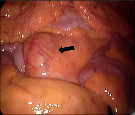
Fig. 2 Laparoscopy shows the tumor (black arrow) located in the mesenteric root.
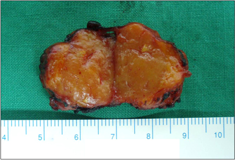
Fig. 3 Gross finding of the resected specimen. The mass shows a 2.5×2 cm in size solid mass with homogenous cut surface.
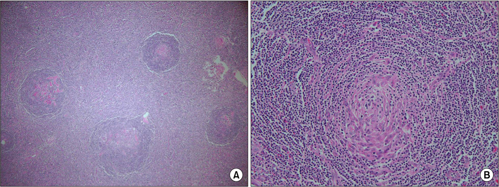
Fig. 4 Microscopic finding of the specimen. (A) Some follicles with marked vascular proliferation and hyalinization. (H&E, ×40). (B) Germinal center with hyalinization and a tight concentric layering of lymphocytes. (onion-skin appearance) (H&E, ×200).
Reference
-
1. Castleman B, Towne VW. Case records of the Massachusetts General Hospital: case no. 40231. N Engl J Med. 1954. 250:1001–1005.2. Choi BJ, Kim KW, An CH, Kim JS, Yoo SJ, Lim KW. Ileal mesenteric Castleman's disease. J Korean Surg Soc. 2005. 69:273–277.3. Hayashi M, Aoshiba K, Shimada M, Izawa Y, Yasui S, Nagai A. Kaposi's sarcoma-associated herpesvirus infection in the lung in multicentric Castleman's disease. Intern Med. 1999. 38:279–282.4. Keller AR, Hochholzer L, Castleman B. Hyaline-vascular and plasma-cell types of giant lymph node hyperplasia of the mediastinum and other locations. Cancer. 1972. 29:670–683.5. Irsutti M, Paul JL, Selves J, Railhac JJ. Castleman disease: CT and MR imaging features of a retroperitoneal location in association with paraneoplastic pemphigus. Eur Radiol. 1999. 9:1219–1221.6. Herrada J, Cabanillas F, Rice L, Manning J, Pugh W. The clinical behavior of localized and multicentric Castleman disease. Ann Intern Med. 1998. 128:657–662.7. Chronowski GM, Ha CS, Wilder RB, Cabanillas F, Manning J, Cox JD. Treatment of unicentric and multicentric Castleman disease and the role of radiotherapy. Cancer. 2001. 92:670–676.8. Williams MD, Eissien FA, Salameh JR, Ailawadi G, Sweeney JF. Laparoscopic approach to the management of intraabdominal unicentric Castleman's disease. Surg Endosc. 2003. 17:1497.9. Jeong CY, Lee YJ, Hong SC, Jung EJ, Choi SK, Joo YT, et al. Castleman's disease in unusual location; plasma cell variant. J Korean Surg Soc. 2005. 68:443–447.
- Full Text Links
-
- Actions
-
Cited
- CITED
-
- Close
- Share
-
- Similar articles
-
- Castleman Disease Arising from IVlesentery: A Case Report
- Mesenteric Castleman's disease(plasma cell type): CT & ultrasound imaging
- Mesenteric Castleman's Disease
- Case Report: Superior Mesenteric Artery Syndrome following Laparoscopic Adjustable Gastric Banding
- MRI findings of castleman disease (Giant lymph node hyperplasia): case report
