Tentorial Dural Arteriovenous Fistula Treated Using Transarterial Onyx Embolization
- Affiliations
-
- 1Department of Radiology, Daejeon St. Mary's Hospital, College of Medicine, The Catholic University of Korea, Seoul, Korea.
- 2Department of Neurosurgery, Daejeon St. Mary's Hospital, College of Medicine, The Catholic University of Korea, Seoul, Korea. hyungjin@catholic.ac.kr
- KMID: 2191383
- DOI: http://doi.org/10.3340/jkns.2015.58.3.276
Abstract
- Tentorial dural arteriovenous fistula (DAVF) is a rare vascular disease, which has high risk of intracranial hemorrhage. We present two cases of tentorial DAVF which were successfully treated with single trial of transarterial embolization using Onyx. We briefly reviewed the types of the tentorial DAVF and strategies of treatment.
Keyword
Figure
-
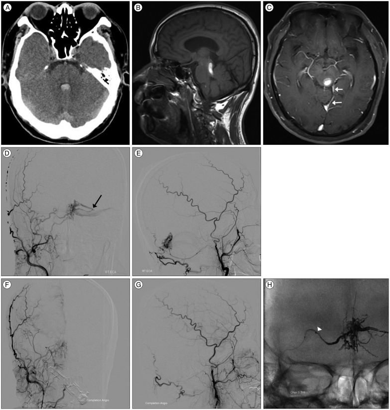
Fig. 1 Case 1. Brain CT (A) and MR imaging (B and C) showed acute hemorrhage at 4th ventricle and left tectum of mid brain. Enhanced axial MR imaging (C) showed prominent vessels near the perimesencephalic cistern and tentorial leaflet (white arrows). Right external carotid angiogram demonstrated a DAVF around torcular fed by meningeal branch of ascending pharyngeal artery, transosseus branch of the right occipital artery and drained into cortical vein of left occiput (black arrow) and basal vein of Rosenthal (D and E). Post-embolization angiogram demonstrated Onyx occupying the retrograde drainage of cortical vein and obliteration of DAVF. Onyx was injected through the transosseus branch of the right occipital artery (arrowhead) (F and G : Right external carotid angiogram. H : Skull AP view).

Fig. 2 Case 2. Brain CT image showed a prominent and tortuous vascular structure around medial tentorium (arrow) (A). Brain MR image showed multiple fine vessel engorgements at both thalamic and putamen regions (B). Angiogram demonstrated recanalization and recruitment from right middle meningeal arteries (C) and left middle meningeal arteries (D and E). The retrograde leptomeningeal drain of infratentorial vermian vein showed dilated and saccular change (arrowhead). Vertebral angiogram also showed supply from posterior choroidal artery (F). After embolization of DAVF through the right middle meningeal artery using Onyx, multiple fistula points and common draining vein was occluded and DAVF was obliterated (G : Right external carotid angiogram. H : Left external carotid angiogram. I : Vertebral angiogram. J : Skull lateral view).
Reference
-
1. Awad IA, Little JR, Akarawi WP, Ahl J. Intracranial dural arteriovenous malformations : factors predisposing to an aggressive neurological course. J Neurosurg. 1990; 72:839–850. PMID: 2140125.
Article2. Borden JA, Wu JK, Shucart WA. A proposed classification for spinal and cranial dural arteriovenous fistulous malformations and implications for treatment. J Neurosurg. 1995; 82:166–179. PMID: 7815143.
Article3. Cognard C, Gobin YP, Pierot L, Bailly AL, Houdart E, Casasco A, et al. Cerebral dural arteriovenous fistulas : clinical and angiographic correlation with a revised classification of venous drainage. Radiology. 1995; 194:671–680. PMID: 7862961.
Article4. Davies MA, Ter Brugge K, Willinsky R, Wallace MC. The natural history and management of intracranial dural arteriovenous fistulae. Part 2 : aggressive lesions. Interv Neuroradiol. 1997; 3:303–311. PMID: 20678361.
Article5. Gruber A, Mazal PR, Bavinzski G, Killer M, Budka H, Richling B. Repermeation of partially embolized cerebral arteriovenous malformations : a clinical, radiologic, and histologic study. AJNR Am J Neuroradiol. 1996; 17:1323–1331. PMID: 8871719.6. Huang Q, Xu Y, Hong B, Li Q, Zhao W, Liu J. Use of onyx in the management of tentorial dural arteriovenous fistulae. Neurosurgery. 2009; 65:287–292. discussion 292-293PMID: 19625907.
Article7. Hwang G, Kang HS, Oh CW, Kwon OK. Surgical obliteration in superior petrosal sinus dural arteriovenous fistula. J Korean Neurosurg Soc. 2011; 49:222–225. PMID: 21607180.
Article8. Jiang C, Lv X, Li Y, Zhang J, Wu Z. Endovascular treatment of high-risk tentorial dural arteriovenous fistulas : clinical outcomes. Neuroradiology. 2009; 51:103–111. PMID: 18989665.
Article9. Lasjaunias P, Chiu M, ter Brugge K, Tolia A, Hurth M, Bernstein M. Neurological manifestations of intracranial dural arteriovenous malformations. J Neurosurg. 1986; 64:724–730. PMID: 3701421.
Article10. Lawton MT, Sanchez-Mejia RO, Pham D, Tan J, Halbach VV. Tentorial dural arteriovenous fistulae : operative strategies and microsurgical results for six types. Neurosurgery. 2008; 62(3 Suppl 1):110–124. discussion 124-125PMID: 18424975.11. Lewis AI, Tomsick TA, Tew JM Jr. Management of tentorial dural arteriovenous malformations : transarterial embolization combined with stereotactic radiation or surgery. J Neurosurg. 1994; 81:851–859. PMID: 7965115.
Article12. Picard L, Bracard S, Islak C, Roy D, Moreno A, Marchal JC, et al. Dural fistulae of the tentorium cerebelli. Radioanatomical, clinical and therapeutic considerations. J Neuroradiol. 1990; 17:161–181. PMID: 2286839.
- Full Text Links
-
- Actions
-
Cited
- CITED
-
- Close
- Share
-
- Similar articles
-
- Dural Arteriovenous Fistula Involving Transverse Sinus: Successful Embolization Using Onyx(R)
- Dural Arteriovenous Fistula Involving an Isolated Sinus Treated Using Transarterial Onyx Embolization
- Hairball-Like Migration of “Onyx Threads” into the Draining Vein during Transarterial Embolization of a Dural Arteriovenous Fistula: A Case Report and Experimental Validation
- A Case of Intraosseous Dural Arteriovenous Fistulas Involving Diploic Vein Treated with Transarterial Onyx Embolization
- Anatomical safety and precaution of transarterial embolization of a falcotentorial dural arteriovenous fistula fed by the artery of Davidoff and Schechter: Case report and review of the literature
