Intraventricular Cavernous Hemangiomas Located at the Foramen of Monro
- Affiliations
-
- 1Department of Neurosurgery, Ilsan Paik Hospital, College of Medicine, Inje University, Goyang, Korea. lbjguni@hanmail.net
- KMID: 2190554
- DOI: http://doi.org/10.3340/jkns.2012.52.2.144
Abstract
- Intraventricular cavernous hemangiomas are uncommon. Among them, those occurred at the foramen of Monro in the third ventricle may be of particular interest because of its rarity, development of hydrocephalus, being differentiated from other brain lesions. We present a rare case of intraventricular cavernous hemangioma at foramen of Monro which was resected through microsurgery and also review the relevant literatures.
Keyword
MeSH Terms
Figure
-

Fig. 1 Computed tomogram (CT) shows enlargement of both lateral ventricles which is caused by 3 cm sized multi-lobular calcified mass with slightly peripheral rim enhancement. It is located at the foramen of Monro in the third ventricle and seems to be close to the left thalamus parenchyme partially (A). Magnetic resonance image (MRI) reveals a relatively well-delineated third ventricular mass with heterogeneous signal intensities in the center of lesion on T1-weighted image (B). Typical peripheral hemosiderin rim of low signal intensity is not delineated on T2-weighted image (C). Minimal peripheral enhancement at the right posterior portion of the mass is seen on enhanced T1-weighted image (D). There is a minimal perilesional edema in the left thalamus on the FLAIR image (E). The mass around foramen of Monro is seen on enhanced sagittal T1-weighted image (F).

Fig. 2 Operative finding shows that the mass with xanthochromic and lobular surface obstructing the foramen of Monro is located in the third ventricle. The left posterior margin of the mass is close to the thalamus.
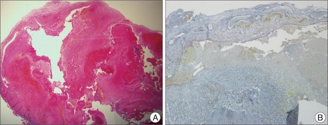
Fig. 3 The histological examinations reveal a large hyalinized vessels filled with organizing thrombus (A). Immunostaining for smooth muscle actin (SMA) is positive in the vessel wall (B).
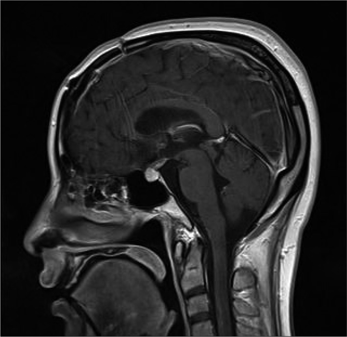
Fig. 4 Post operative sagittal MRI shows no evidence of residual mass and improved hydrocephalus.
Reference
-
1. Chen CL, Leu CH, Jan YJ, Shen CC. Intraventricular cavernous hemangioma at the foramen of Monro : case report and literature review. Clin Neurol Neurosurg. 2006; 108:604–609. PMID: 15916846.
Article2. Clatterbuck RE, Eberhart CG, Crain BJ, Rigamonti D. Ultrastructural and immunocytochemical evidence that an incompetent blood-brain barrier is related to the pathophysiology of cavernous malformations. J Neurol Neurosurg Psychiatry. 2001; 71:188–192. PMID: 11459890.
Article3. Katayama Y, Tsubokawa T, Maeda T, Yamamoto T. Surgical management of cavernous malformations of the third ventricle. J Neurosurg. 1994; 80:64–72. PMID: 8271024.
Article
- Full Text Links
-
- Actions
-
Cited
- CITED
-
- Close
- Share
-
- Similar articles
-
- Erratum: Intraventricular Cavernous Hemangiomas Located at the Foramen of Monro
- A Method for the Localization of Normal Foramen of Monro in Korean
- Subcutaneous Cavernous and Capillary Hemangiomas of the Breast: Radiologic-Pathological Correlation
- A Case of Extradural Cavernous Hemangioma with Reuptured Disc
- A case of esophageal cavernous hemangioma
