J Korean Assoc Oral Maxillofac Surg.
2011 Feb;37(1):49-53. 10.5125/jkaoms.2011.37.1.49.
Comparative study of removal torque of 3 different hydroxyapatite coated implants in the femur of rabbits
- Affiliations
-
- 1Division of Oral and Maxillofacial, Department of Dentistry, Daegu Catholic University Medical Center, Daegu, Korea. dssohn@cu.ac.kr
- KMID: 2189806
- DOI: http://doi.org/10.5125/jkaoms.2011.37.1.49
Abstract
- INTRODUCTION
This study compared the strength of osseointegration as determined by the resistance to reverse torque rotation of three different hydroxyapatite coated implants in the rabbit femur model.
MATERIALS AND METHODS
Three hydroxyapatite coated implants (HAPTITE), Tapered Screw-Vent (TSV) and BioTite-H - were used. A total of 40 implants were placed in the femur of 20 adult male rabbits. The animals were divided into two groups. In group A (n=10); one HAPTITE was placed into each right femur and one TSV was placed into each left femur. In group B (n=10); one HAPTITE was placed into each right femur and one BioTite-H was placed into each left femur. Five rabbits of each group were sacrificed at 4 and 8 weeks. The implants were removed by reverse torque rotation using a digital torque-measuring device. A total of 40 implants in 20 rabbits were used for the removal torque measurements.
RESULTS
In the Group A, 4 weeks after implant placement, the mean removal torque for the HAPTITE and TSV was 70.7+/-31.6 N cm and 28.9+/-15.1 N cm, respectively. Eight weeks after implant placement, the mean removal torque for the HAPTITE and TSV was 87.9+/-26.2 N cm and 54.9+/-22.4 N cm, respectively. In the Group B, 4 weeks after implant placement, the mean removal torque for the HAPTITE and BioTite-H was 58.0+/-29.6 N cm and 37.7+/-14.1 N cm, respectively. Eight weeks after implant placement, the mean removal torque for the HAPTITE and BioTite-H was 91.4+/-47.1 N cm and 30.8+/-9.8 N cm. HAPTITE showed a higher removal torque than the other implants.
CONCLUSION
These results suggest that HAPTITE increases the strength of osseointegration significantly as determined by the resistance to reverse torque rotation.
Keyword
MeSH Terms
Figure
-
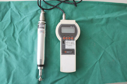
Fig. 1. Digital torque meter used to measure the peak value of resistance to reverse torque rotation in N cm.
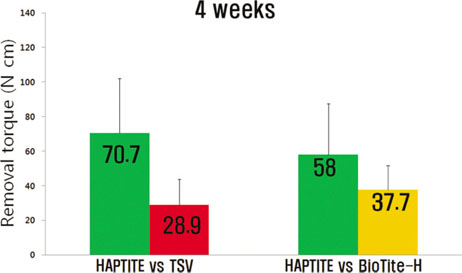
Fig. 2. Mean removal torque values (N cm) of implants 4 weeks after implantation.(TSV: Tapered Screw-Vent)
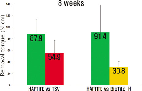
Fig. 3. Mean removal torque values (N cm) of implants 8 weeks after implantation.(TSV: Tapered Screw-Vent)
Cited by 1 articles
-
Hydroxyapatite-coated implant: Clinical prognosis assessment via a retrospective follow-up study for the average of 3 years
Jun-Hong Jung, Sang-Yun Kim, Yang-Jin Yi, Bu-Kyu Lee, Young-Kyun Kim
J Adv Prosthodont. 2018;10(2):85-92. doi: 10.4047/jap.2018.10.2.85.
Reference
-
1. Brånemark PI, Adell R, Breine U, Hansson BO, Hutton JE, Lindstro¨m J, Ohlsson A. Intra-osseous anchorage of dental prostheses. I. Experimental studies. Scand J Plast Reconstr Surg. 1969; 3:81–100.
Article2. Hutton JE, Heath MR, Chai JY, Harnett J, Jemt T, Johns RB, et al. Factors related to success and failure rates at 3-year follow-up in a multicenter study of overdentures supported by Brånemark implants. Int J Oral Maxillofac Implants. 1995; 10:33–42.3. Albrektsson T, Zarb G, Worthington P, Eriksson AR. The long-term efficacy of currently used dental implants: a review and proposed criteria of success. Int J Oral Maxillofac Implants. 1986; 1:11–25.4. Shirakura M, Fujii N, Ohnishi H, Taguchi Y, Ohshima H, Nomura S, et al. Tissue response to titanium implantation in the rat maxilla, with special reference to the effects of surface conditions on bone formation. Clin Oral Implants Res. 2003; 14:687–696.5. Franchi M, Fini M, Martini D, Orsini E, Leonardi L, Ruggeri A, et al. Biological fixation of endosseous implants. Micron. 2005; 36:665–671.6. Predecki P, Auslaender BA, Stephan JE, Mooney VL, Stanitski C. Attachment of bone to threaded implants by ingrowth and mechanical interlocking. J Biomed Mater Res. 1972; 6:401–412.7. Cook SD, Kay JF, Thomas KA, Jarcho M. Interface mechanics and histology of titanium and hydroxylapatite-coated titanium for dental implant applications. Int J Oral Maxillofac Implants. 1987; 2:15–22.8. Vercaigne S, Wolke JG, Naert I, Jansen JA. Histomorphometrical and mechanical evaluation of titanium plasma-spray-coated implants placed in the cortical bone of goats. J Biomed Mater Res. 1998; 41:41–48.
Article9. Buser D, Schenk RK, Steinemann S, Fiorellini JP, Fox CH, Stich H. Influence of surface characteristics on bone integration of titanium implants. A histomorphometric study in miniature pigs. J Biomed Mater Res. 1991; 25:889–902.
Article10. Cochran DL, Schenk RK, Lussi A, Higginbottom FL, Buser D. Bone response to unloaded and loaded titanium implants with a sandblasted and acid-etched surface: a histometric study in the canine mandible. J Biomed Mater Res. 1998; 40:1–11.11. Søballe K, Hansen ES, Brockstedt-Rasmussen H, Pedersen CM, Bünger C. Hydroxyapatite coating enhances fixation of porous coated implants. A comparison in dogs between press fit and noninterference fit. Acta Orthop Scand. 1990; 61:299–306.12. Block MS, Kent JN, Kay JF. Evaluation of hydroxylapatite-coated titanium dental implants in dogs. J Oral Maxillofac Surg. 1987; 45:601–607.
Article13. Wennerberg A, Albrektsson T, Andersson B, Krol JJ. A histomorphometric and removal torque study of screw-shaped titanium implants with three different surface topographies. Clin Oral Implants Res. 1995; 6:24–30.
Article14. Kumar M, Dasarathy H, Riley C. Electrodeposition of brushite coatings and their transformation to hydroxyapatite in aqueous solutions. J Biomed Mater Res. 1999; 45:302–310.15. Baker D, London RM, O\'Neal R. Rate of pull-out strength gain of dual-etched titanium implants: a comparative study in rabbits. Int J Oral Maxillofac Implants. 1999; 14:722–728.16. Masuda T, Yliheikkilä PK, Felton DA, Cooper LF. Generalizations regarding the process and phenomenon of osseointegration. Part I. In vivo studies. Int J Oral Maxillofac Implants. 1998; 13:17–29.17. Cooper LF, Masuda T, Yliheikkilä PK, Felton DA. Generalizations regarding the process and phenomenon of osseointegration. Part II. In vitro studies. Int J Oral Maxillofac Implants. 1998; 13:163–174.18. Rich A, Harris AK. Anomalous preferences of cultured macrophages for hydrophobic and roughened substrata. J Cell Sci. 1981; 50:1–7.19. Johansson C, Albrektsson T. Integration of screw implants in the rabbit: a 1-year follow-up of removal torque of titanium implants. Int J Oral Maxillofac Implants. 1987; 2:69–75.20. Sakakura CE, Margonar R, Holzhausen M, Nociti FH Jr, Alba RC Jr, Marcantonio E Jr. Influence of cyclosporin A therapy on bone healing around titanium implants: a histometric and biomechanic study in rabbits. J Periodontol. 2003; 74:976–981.21. Sennerby L, Thomsen P, Ericson LE. A morphometric and biomechanic comparison of titanium implants inserted in rabbit cortical and cancellous bone. Int J Oral Maxillofac Implants. 1992; 7:62–71.22. Ivanoff CJ, Widmark G, Johansson C, Wennerberg A. Histologic evaluation of bone response to oxidized and turned titanium micro-implants in human jawbone. Int J Oral Maxillofac Implants. 2003; 18:341–348.23. Roberts WE, Garreto LP. Bone physiology and metabolism. In: Misch CE, editor. Contemporary implant dentistry. 2nd ed. St. Louis: Mosby Co.; 1999. pp. 234-235.24. Misch CM. Hydroxylapatite-coated implants. Design considerations and clinical parameters. N Y State Dent J 1993;59:36-41. Erratum in. N Y State Dent J. 1993; 59:12.
Article25. Davies JE, Lowenberg B, Shiga A. The bone-titanium interface in vitro. J Biomed Mater Res. 1990; 24:1289–1306.
Article26. Yang Y, Bumgardner JD, Cavin R, Carnes DL, Ong JL. Osteoblast precursor cell attachment on heat-treated calcium phosphate coatings. J Dent Res. 2003; 82:449–453.
Article27. Clèries L, Fernández-Pradas JM, Sardin G, Morenza JL. Dissolution behaviour of calcium phosphate coatings obtained by laser ablation. Biomaterials. 1998; 19:1483–1487.
Article
- Full Text Links
-
- Actions
-
Cited
- CITED
-
- Close
- Share
-
- Similar articles
-
- Histomorphometric and Removal Torque Values Comparision of Rough Surface Titanium Implants
- The evaluation of the removal torque and the histomorphometry of the Ca-P coating surface in rabbit tibia
- EFFECTS OF THE ION BEAM ASSISTED DEPOSITION OF HYDROXYAPATITE ON OSSEOINTEGRATION OF THE ENDOSSEOUS IMPLANTS IN RABBIT TIBIAE
- Comparison of removal torque of saline-soaking RBM implants and RBM implants in rabbit tibias
- The Biological Effects of Calcium Phosphate Coated Implant for Osseointegration in Beagle Dogs
