J Korean Med Assoc.
2005 Jul;48(7):628-633. 10.5124/jkma.2005.48.7.628.
New Strategy of Ocular Surface Disease: Ocular Surface Reconstruction Using Amniotic Membrane and Limbal Stem Cell Transplantation
- Affiliations
-
- 1Department of Ophthalmology, Dong-A University College of Medicine and Hospital, Korea. wcpark@dau.ac.kr
- KMID: 2183588
- DOI: http://doi.org/10.5124/jkma.2005.48.7.628
Abstract
- Amniotic membrane is the innermost layer of the placenta and consists of a thick basement membrane and an avascular stromal matrix. Amniotic membrane transplantation facilitates rapid healing with recovery of a normal epithelial phenotype in the epithelium, and reduces inflammation, vascularization, and scarring in the stroma. Amniotic membrane has two major role, basement membrane and cocktail of cytokines. Amniotic membrane as a native matrix can be used as a graft to restore conjunctival surfaces and corneal surfaces suffering from intractable corneal disease or limbal stem cell deficiency. To restore limbal stem cell deficiency, the source of stem cell has been introduced the autograft transplantation, allograft transplantation, ex vivo expansion and in vivo expansion.
Keyword
MeSH Terms
Figure
-

Figure 1

Figure 2
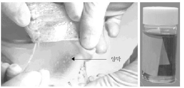
Figure 3
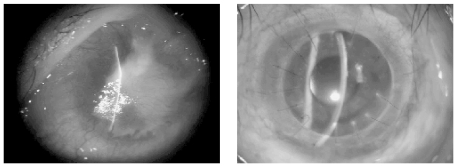
Figure 4
- Full Text Links
-
- Actions
-
Cited
- CITED
-
- Close
- Share
-
- Similar articles
-
- Ocular Surface Reconstruction with Amniotic Membrane Transplantation in Pterygium
- Transplantation of in vivo Cultivated Limbal Corneal Epithelial Cells with Total Limbal Stem Cell Deficiency
- The Effect of Limbal Transplantation & Cyclosporine A for Chemically Damaged Rabbit Cornea
- Living Related Conjunctival Limbal Allograft
- Amniotic Membrane Transplanted in Conjunctiva as a Mesenchymal Stem Cells Carrier for Limbal Stem Cell Deficiency
