Lateral Decubitus Positioning Stereotactic Vacuum-Assisted Breast Biopsy with True Lateral Mammography
- Affiliations
-
- 1Breast Cancer Center, Pusan National University Hospital, Busan, Korea.
- 2Department of Surgery, Pusan National University Hospital, Busan, Korea. bytae@pusan.ac.kr
- 3Department of Pathology, Pusan National University Hospital, Busan, Korea.
- 4Department of Radiology, Yangsan Pusan National University Hospital, Pusan National University School of Medicine, Yangsan, Korea.
- KMID: 2175645
- DOI: http://doi.org/10.4048/jbc.2011.14.1.64
Abstract
- Stereotactic vacuum-assisted breast biopsy (VAB) has been used to evaluate microcalcifications or non-palpable breast lesions on mammography. Although stereotactic VAB is usually performed in a prone or upright position, an expensive prone table is necessary and vasovagal reactions often occur during the procedure. For these reasons, the lateral decubitus position can be applied for stereotactic VAB, and true lateral mammography can be used to detect the lesion. We report on 15 cases of lateral decubitus positioning for stereotactic VAB with true lateral mammography for non-palpable breast lesions or microcalcifications. The mean procedure time was approximately 30.1 minutes, and no complications occurred during the procedures. Fourteen cases had benign breast lesions and one case had a ductal carcinoma in situ. The lateral decubitus stereotactic VAB with true lateral mammography can be applied for microcalcifications or non-palpable breast lesions and helps to minimize anxiety and vasovagal reactions in patients.
MeSH Terms
Figure
-
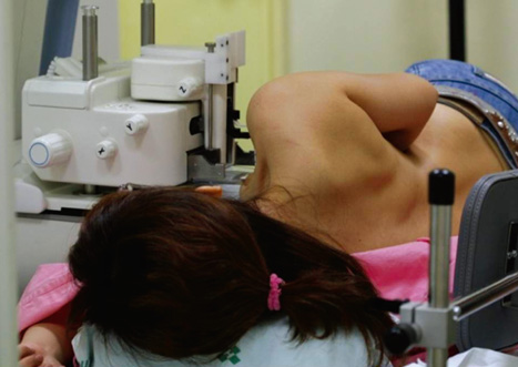
Figure 1 Patient laid on the stereotactic vacuum-assisted breast biopsy table in the lateral decubitus position with the breast lesion upside.

Figure 2 A scout 15° paired stereotactic mammography view. The targeted microcalcification is seen below the probe.
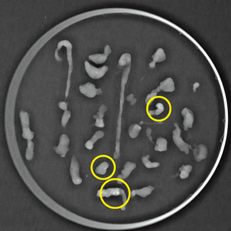
Figure 3 Specimen mammography shows a microcalcification in three separate specimens (yellow circles).
Cited by 2 articles
-
Absence of Residual Microcalcifications in Atypical Ductal Hyperplasia Diagnosed via Stereotactic Vacuum-Assisted Breast Biopsy: Is Surgical Excision Obviated?
Inyoung Youn, Min Jung Kim, Hee Jung Moon, Eun-Kyung Kim
J Breast Cancer. 2014;17(3):265-269. doi: 10.4048/jbc.2014.17.3.265.Clinical Experience of Ultrasound-Guided, Vacuum-Assisted Breast Biopsy for Mammographic Microcalcifications: Combination with Wire Localization
SeungSang Ko, Man Sik Shin, Ki Won Chun, Kang Young Rhee, Heeboong Park
J Surg Ultrasound. 2018;5(2):53-60. doi: 10.46268/jsu.2018.5.2.53.
Reference
-
1. Schmidt RA. Stereotactic breast biopsy. CA Cancer J Clin. 1994. 44:172–191.
Article2. Ohsumi S, Takashima S, Aogi K, Ishizaki M, Mandai K. Breast biopsy for mammographically detected non-palpable lesions using a vacuum-assisted biopsy device (Mammotome) and an upright-type stereotactic mammography unit. Jpn J Clin Oncol. 2001. 31:527–531.
Article3. Sim LS, Kei PL. Upright stereotactic vacuum-assisted needle biopsy of suspicious breast microcalcifications. J Med Imaging Radiat Oncol. 2008. 52:358–364.
Article4. Rovera F, Dionigi G, Marelli M, Ferrari A, Limonta G, Corben AD, et al. Breast cancer diagnosis: the role of stereotactic vacuum-assisted aspiration biopsy. Int J Surg. 2008. 6:Suppl 1. S104–S108.
Article5. Nisbet AP, Borthwick-Clarke A, Scott N. 11-gauge vacuum assisted directional biopsy of breast calcifications, using upright stereotactic guidance. Eur J Radiol. 2000. 36:144–146.
Article6. Wunderbaldinger P, Wolf G, Turetschek K, Helbich TH. Comparison of sitting versus prone position for stereotactic large-core breast biopsy in surgically proven lesions. AJR Am J Roentgenol. 2002. 178:1221–1225.
Article7. Welle GJ, Clark M, Loos S, Pauls D, Warden D, Sheffield M, et al. Stereotactic breast biopsy: recumbent biopsy using add-on upright equipment. AJR Am J Roentgenol. 2000. 175:59–63.8. Yu YH, Liang C, Yuan XZ. Diagnostic value of vacuum-assisted breast biopsy for breast carcinoma: a meta-analysis and systematic review. Breast Cancer Res Treat. 2010. 120:469–479.
Article9. Kettritz U, Rotter K, Schreer I, Murauer M, Schulz-Wendtland R, Peter D, et al. Stereotactic vacuum-assisted breast biopsy in 2874 patients: a multicenter study. Cancer. 2004. 100:245–251.
Article10. Apesteguía L, Mellado M, Sáenz J, Cordero JL, Repáraz B, De Miguel C. Vacuum-assisted breast biopsy on digital stereotaxic table of nonpalpable lesions non-recognisable by ultrasonography. Eur Radiol. 2002. 12:638–645.
Article11. Choo KS, Kwak HS, Tae Bae Y, Lee JY, Lee SJ, Seo HI, et al. The value of a combination of wire localization and ultrasound-guided vacuum-assisted breast biopsy for clustered microcalcifications. Breast. 2008. 17:611–616.
Article12. Hung WK, Lam HS, Lau Y, Chan CM, Yip AW. Diagnostic accuracy of vacuum-assisted biopsy device for image-detected breast lesions. ANZ J Surg. 2001. 71:457–460.
Article13. Zagouri F, Sergentanis TN, Nonni A, Koulocheri D, Fotou M, Panopoulou E, et al. Vacuum-assisted breast biopsy: the value and limitations of cores with microcalcifications. Pathol Res Pract. 2007. 203:563–566.
Article14. Yom CK, Moon BI, Choe KJ, Choi HY, Park YL. Long-term results after excision of breast mass using a vacuum-assisted biopsy device. ANZ J Surg. 2009. 79:794–798.
Article15. Fine RE, Whitworth PW, Kim JA, Harness JK, Boyd BA, Burak WE Jr. Low-risk palpable breast masses removed using a vacuum-assisted hand-held device. Am J Surg. 2003. 186:362–367.
Article
- Full Text Links
-
- Actions
-
Cited
- CITED
-
- Close
- Share
-
- Similar articles
-
- Stereotactic vacuum-assisted breast biopsy under lateral decubitus position
- The Clinical Utility of a Adding Lateral Approach to Conventional Vertical Approach for Prone Stereotactic Vacuum-Assisted Breast Biopsy
- Diagnosis of Non-palpable Breast Lesions with Microcalcification by Upright Add-on Type Stereotactic Vacuum-assisted Biopsy
- The Efficacy of Stereotactic Vacuum-assisted Biopsy and Needle Localization Vacuum-assisted Biopsy for Diagnosing Breast Microcalcification
- Mammography-Guided Interventional Procedure
