Establishment and Characterization of Human Lymphatic Endothelial Cell in Breast Cancer Patients Undergoing Sentinel Lymphadenectomy
- Affiliations
-
- 1Department of Surgery and Surgical Oncology and Project Research Division, Research Center for Genomic Medicine, Saitama Medical University, Saitama, Japan. aktake@iuhw.ac.jp
- KMID: 2175195
- DOI: http://doi.org/10.4048/jbc.2009.12.2.61
Abstract
- Recently remarkable progress has been made in the understanding of lymphangiogenesis due to the availability of specific biomarkers. A sentinel lymph node (SLN) biopsy has already been established as a common surgical procedure, and the clinical usefulness of an SLN biopsy has been confirmed in patients with various types of cancer. In this study, a novel method has been successfully developed for the isolation of anatomically defined lymphatic endothelial cells (LECs) from human sentinel lymphatic channels during an SLN biopsy in breast cancer patients by the use of collagenase II digestion. The isolated cells were cultured in EGM-2MV media with 10% fetal bovine serum (FBS) under hypoxic conditions in an atmosphere of 5% O2, 5% CO2 and 90% N2 at 37degrees C. The cultured cells exhibited a monolayer with a cobblestone appearance. Immunofluorescence analysis using selective lymphangiogenesis markers showed expression of vascular endothelial growth factor receptor 3 (VEGFR-3), prospero homeobox 1 (Prox-1) and Podoplanin. Newly established LECs showed expressions of vascular endothelial growth factor (VEGF) family members except VEGF-D and corresponding VEGF receptors by the use of conventional RT-PCR. Treatment with VEGF-C156S (500 ng/mL) apparently induced phosphorylation of the protein kinase Akt, MAPK and JNK in human isolated LECs as determined by Western blot analysis. Peak induction of Akt, MAPK and JNK occurred at 10 to 15 minutes after incubation of the isolated LECs with VEGF-C156S. The use of the MTS cell proliferation assay showed a significant increase in the growth of human lymphatic endothelial cells with VEGF-C156S treatment. The effects of VEGF-C156S (500 ng/mL) on proliferation activity was significantly with 3% FBS condition alone (MTS score: 0.54+/-0.0104, n=3) and a 3% FBS+VEGF-C156S (MTS score: 0.572+/-0.0061, n=3) condition. The isolated and enzymatic digested method adopted for the culture of human LECs is simple and useful for the investigation of the cellular, molecular and genomic properties of LECs.
MeSH Terms
-
Atmosphere
Biomarkers
Biopsy
Blotting, Western
Breast
Breast Neoplasms
Cell Proliferation
Cells, Cultured
Collagenases
Digestion
Endothelial Cells
Fluorescent Antibody Technique
Genes, Homeobox
Humans
Lymph Node Excision
Lymph Nodes
Lymphangiogenesis
Nitriles
Phosphorylation
Protein Kinases
Pyrethrins
Receptors, Vascular Endothelial Growth Factor
Vascular Endothelial Growth Factor A
Vascular Endothelial Growth Factor D
Vascular Endothelial Growth Factor Receptor-3
Collagenases
Nitriles
Protein Kinases
Pyrethrins
Receptors, Vascular Endothelial Growth Factor
Vascular Endothelial Growth Factor A
Vascular Endothelial Growth Factor D
Vascular Endothelial Growth Factor Receptor-3
Figure
-
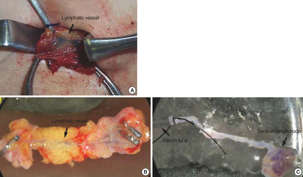
Figure 1 (A) Image of lymphatic vessel containing blue dye in sentinel lymphadenectomy. (B) A representative micrograph of isolated segment of human afferent lymphatic vessel treated by micro-clips at both ends. (C) A representative micrograph of the afferent lymphatic vessel cannulated centripetally with a sterile silicon tube.
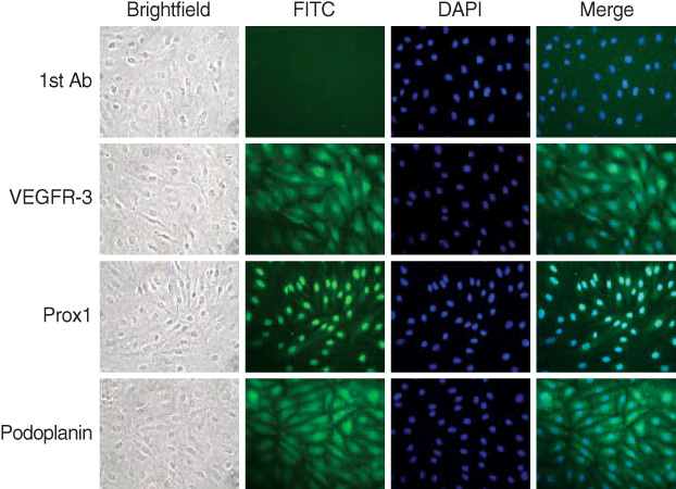
Figure 2 Representative micrographs of immunohistochemical images of VEGFR-3, Prox-1 and Podoplanin in the cultured lymphatic endothelial cells in the collecting lymph vessels (×400).
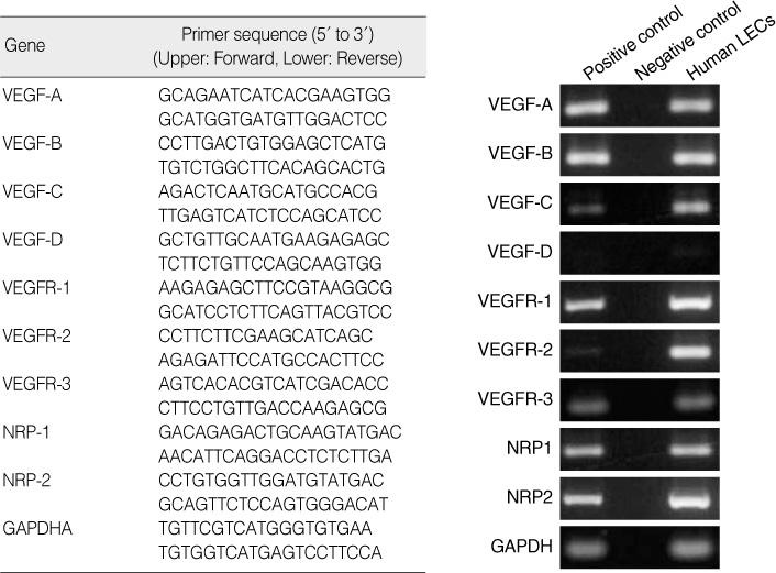
Figure 3 Expression of VEGF families and their receptors by the RT-PCR in our isolated lymphatic endothelial cells. LECs=lymphatic endothelial cells.
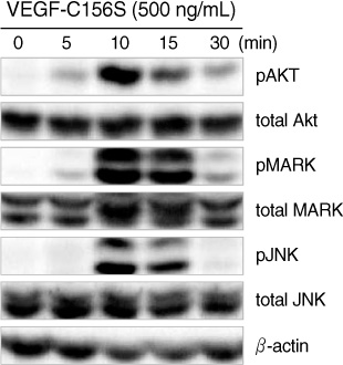
Figure 4 Activated and total Akt, MAPK and JNK levels in protein extracted from human lymphatic endothelial cells treated with VEGF-C156S (500 ng/mL) for the indicated times.
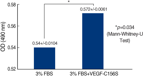
Figure 5 Effects of VEGF-C156S (500 ng/mL) on the proliferation activity of cultured human lymphatic endothelial cells using MTS assay.
Reference
-
1. Ichikura T, Morita D, Uchida T, Okura E, Majima T, Ogawa T, et al. Sentinel node concept in gastric carcinoma. World J Surg. 2002. 26:318–322.
Article2. Hayashi H, Ochiai T, Mori M, Karube T, Suzuki T, Gunji Y, et al. Sentinel lymph node mapping for gastric cancer using a dual procedure with dye- and gamma probe-guided techniques. J Am Coll Surg. 2003. 196:68–74.
Article3. Takeda A, Iseki H, Otani Y, Takeuchi H, Ichioka S, Kawai Y, et al. Lymphatic mapping and lymphatic endothelial cell isolation in colorectal cancer patients. Asia Pac J Clin Oncol. 2006. 2:167–174.
Article4. Detmar M, Hirakawa S. The formation of lymphatic vessels and its importance in the setting of malignancy. J Exp Med. 2002. 196:713–718.
Article5. Mandriota SJ, Jussila L, Jeltsch M, Compagni A, Baetens D, Prevo R, et al. Vascular endothelial growth factor-C-mediated lymphangiogenesis promotes tumour metastasis. EMBO J. 2001. 20:672–682.
Article6. Takeuchi H, Takeda A, Iseki H, Shigekawa T, Takahashi N, Andoh T, et al. Isolation and characterization of human lymphatic endothelial cell from primary breast cancer patients. 2007. In : 66th Annual Meeting of the Japanese Cancer Association Program; 144.7. Mizuno R, Yokoyama Y, Ono N, Ikomi F, Ohhashi T. Establishment of rat lymphatic Endothelial cell line. Microcirculation. 2003. 10:127–131.
Article8. Garrafa E, Alessandri G, Benetti A, Turetta D, Corradi A, Cantoni AM, et al. Isolation and characterization of lymphatic microvascular endothelial cells from human tonsils. J Cell Physiol. 2006. 207:107–113.
Article9. Kawai Y, Minami T, Fujimori M, Hosaka K, Mizuno R, Ikomi F, et al. Characterization and microarray analysis of genes in human lymphatic endothelial cells from patients with breast cancer. Lymphat Res Biol. 2007. 5:115–126.
Article10. Morton DL, Wen DR, Wong JH, Economou JS, Cagle LA, Storm FK, et al. Technical details of intraoperative lymphatic mapping for early stage melanoma. Arch Surg. 1992. 127:392–399.
Article11. Bilchik AJ, Saha S, Wiese D, Stonecypher JA, Wood TF, Sostrin S, et al. Molecular staging of early colon cancer on the basis of sentinel node analysis: a multicenter phase II trial. J Clin Oncol. 2001. 19:1128–1136.
Article12. Paramo JC, Summerall J, Wilson C, Cabral A, Willis I, Wodnicki H, et al. Intraoperative sentinel lymph node mapping in patients with colon cancer. Am J Surg. 2001. 182:40–43.
Article13. Saha S, Dan AG, Beutler T, Schochet E, Badin J, Branigan T, et al. Sentinel lymph node mapping technique in colon cancer. Semin Oncol. 2004. 31:374–381.
Article
- Full Text Links
-
- Actions
-
Cited
- CITED
-
- Close
- Share
-
- Similar articles
-
- Sentinel Lymph Node Biopsy in Breast Cancer: A Clinical Review and Update
- Sentinel Lymph Node Imaging in Breast Cancer
- Validation and Controversy of Sentinel Node Biopsy for Breast Cancer
- Surgical treatment of early ovarian cancer with compartmental resection of regional lymphatic network and indocyanine-green-guided targeted compartmental lymphadenectomy (TCL, paraaortic part)
- Anatomy Versus Physiology: Is Breast Lymphatic Drainage to the Internal Thoracic (Internal Mammary) Lymphatic System Clinically Relevant?
