Diagnostic performance of cone-beam computed tomography on detection of mechanically-created artificial secondary caries
- Affiliations
-
- 1Division of Oral and Maxillofacial Radiology, Department of Oral Biology and Diagnostic Sciences, Faculty of Dentistry, Chiang Mai University, Chiang Mai, Thailand. arnon_cha903@hotmail.com
- KMID: 2167398
- DOI: http://doi.org/10.5624/isd.2011.41.4.143
Abstract
- PURPOSE
The aim of this study was to compare the diagnostic accuracy of cone-beam computed tomography (CBCT) images and bitewing images in detection of secondary caries.
MATERIALS AND METHODS
One hundred and twenty proximal slots of Class II cavities were randomly prepared on human premolar and molar teeth, and restored with amalgam (n=60) and composite resin (n=60). Then, artificial secondary caries lesions were randomly created using round steel No. 4 bur. The teeth were radiographed with a conventional bitewing technique and two CBCT systems; Pax-500ECT and Promax 3D. All images were evaluated by five observers. The area under the receiver operating characteristic (ROC) curve (Az) was used to evaluate the diagnostic accuracy. Significant difference was tested using the Friedman test (p value<0.05).
RESULTS
The mean Az values for bitewing, Pax-500ECT, and Promax 3D imaging systems were 0.882, 0.995, and 0.978, respectively. Significant differences were found between the two CBCT systems and film (p=0.007). For CBCT systems, the axial plane showed the greatest Az value.
CONCLUSION
Based on the design of this study, CBCT images were better than bitewing radiographs in detection of secondary caries.
Keyword
MeSH Terms
Figure
-
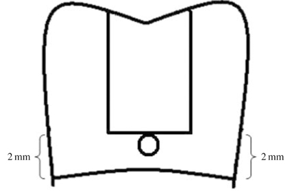
Fig. 1 The illustration shows artificial caries prepared at the gingival floor and sealed with pink wax.
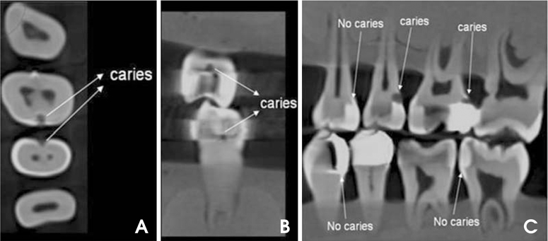
Fig. 2 Examples of CBCT images demonstrates either secondary caries present or absent. A. Axial plane. B. Coronal plane. C. Sagittal plane.
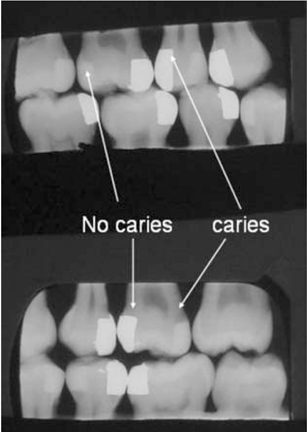
Fig. 3 Examples of bitewing images demonstrate either secondary caries present or absent
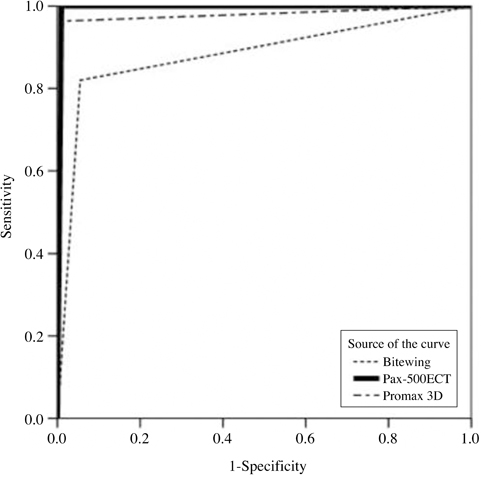
Fig. 4 ROC curves represent pooled radiographic scores for five observers diagnosing secondary caries in connection with imaging modalities.
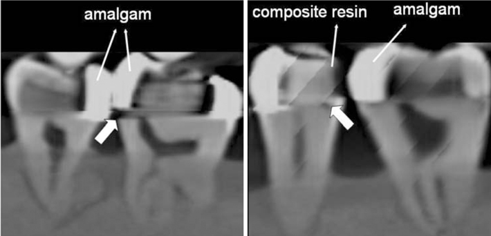
Fig. 5 Examples of CBCT images in sagittal plane in which misinterpretation of secondary caries occurred. Thick arrows point at the surfaces that gave false positive results.
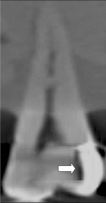
Fig. 6 The CBCT image in the sagittal plane shows the artifacts (arrow) from the adjacent amalgam restoration that could be misinterpreted as secondary or remnant caries.
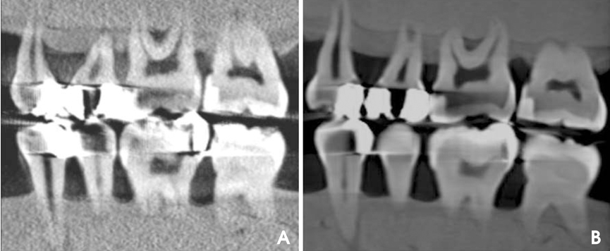
Fig. 7 The Promax 3D system CBCT image (A) demonstrates a greater amount of artifacts than that of the Pax-500ECT system (B).
Reference
-
1. Scarfe WC, Farman AG. White SC, Pharoah MJ, editors. Cone-beam computed tomography. Oral radiology: principle and interpretation. 2009. 6th ed. St. Louis: Mosby;225–242.
Article2. Akdeniz B, Gröndahl H, Magnusson B. Accuracy of proximal caries depth measurements: comparison between limited cone beam computed tomography, storage phosphor and film radiography. Caries Res. 2006. 40:202–207.
Article3. Tsuchida R, Araki K, Okano T. Evaluation of a limited cone-beam volumetric imaging system: comparison with film radiography in detecting incipient proximal caries. Oral Surg Oral Med Oral Pathol Oral Radiol Endod. 2007. 104:412–416.
Article4. Haiter-Neto F, Wenzel A, Gotfredsen E. Diagnostic accuracy of cone beam computed tomography scans compared with intraoral image modalities for detection of caries lesions. Dentomaxillofac Radiol. 2008. 37:18–22.
Article5. Young SM, Lee JT, Hodges RJ, Chang TL, Elashoff DA, White SC. A comparative study of high-resolution cone beam computed tomography and charge-coupled device sensors for detecting caries. Dentomaxillofac Radiol. 2009. 38:445–451.
Article6. Qu X, Li G, Zhang Z, Ma X. Detection accuracy of in vitro approximal caries by cone beam computed tomography images. Eur J Radiol. 2011. 79:e24–e27.
Article7. Kayipmaz S, Sezgin OS, Saricaoğlu ST, Can G. An in vitro comparison of diagnostic abilities of conventional radiography, storage phosphor, and cone beam computed tomography to determine occlusal and approximal caries. Eur J Radiol. 2011. 80:478–482.
Article8. Zhang ZL, Qu XM, Li G, Zhang ZY, Ma XC. The detection accuracies for proximal caries by cone-beam computerized tomography, film, and phosphor plates. Oral Surg Oral Med Oral Pathol Oral Radiol Endod. 2011. 111:103–108.
Article9. Kamburoğlu K, Murat S, Yüksel SP, Cebeci AR, Paksoy CS. Occlusal caries detection by using a cone-beam CT with different voxel resolutions and a digital intraoral sensor. Oral Surg Oral Med Oral Pathol Oral Radiol Endod. 2010. 109:e63–e69.
Article10. Kidd EA. Diagnosis of secondary caries. J Dent Educ. 2001. 65:997–1000.
Article11. Mjör IA, Toffenetti F. Secondary caries: a literature review with case reports. Quintessence Int. 2000. 31:165–179.12. Imperiano MT, Khoury HJ, Pontual ML, Montes MA, Silveira MM. Comparative radiopacity of four low-viscosity composites. Braz J Oral Sci. 2007. 6:1278–1282.13. Nair MK, Tyndall DA, Ludlow JB, May K. Tuned aperture computed tomography and detection of recurrent caries. Caries Res. 1998. 32:23–30.
Article14. Espelid I, Tveit AB, Erickson RL, Keck SC, Glasspoole EA. Radiopacity of restorations and detection of secondary caries. Dent Mater. 1991. 7:114–117.
Article15. Espelid I, Tveit AB. Diagnosis of secondary caries and crevices adjacent to amalgam. Int Dent J. 1991. 41:359–364.16. Matteson SR, Phillips C, Kantor ML, Leinedecker T. The effect of lesion size, restorative material, and film speed on the detection of recurrent caries. Oral Surg Oral Med Oral Pathol. 1989. 68:232–237.
Article17. Kang B-C, Farman AG, Scarfe WC, Goldsmith LJ. Mechanical defects in dental enamel vs. natural dental caries: observer differentiation using Ektaspeed Plus film. Caries Res. 1996. 30:156–162.
Article18. Nair M, Tyndall D, Ludlow J, May K, Ye F. The effects of restorative material and location on the detection of simulated recurrent caries. A comparison of dental film, direct digital radiography and tuned aperture computed tomography. Dentomaxillofac Radiol. 1998. 27:80–84.
Article19. Grossman ES, Matejka JM. Histological features of artificial secondary caries adjacent to amalgam restorations. J Oral Rehabil. 1999. 26:737–744.
Article20. Hassan B, Metska ME, Ozok AR, van der Stelt P, Wesselink PR. Detection of vertical root fractures in endodontically treated teeth by a cone beam computed tomography scan. J Endod. 2009. 35:719–722.
Article21. Sanders MA, Hoyjberg C, Chu CB, Leggitt VL, Kim JS. Common orthodontic appliances cause artifacts that degrade the diagnostic quality of CBCT Images. J Calif Dent Assoc. 2007. 35:850–857.22. Cevidanes LH, Tucker S, Styner M, Kim H, Chapuis J, Reyes M, et al. Three-dimensional surgical simulation. Am J Orthod Dentofacial Orthop. 2010. 138:361–371.
Article23. Razavi T, Palmer RM, Davies J, Wilson R, Palmer PJ. Accuracy of measuring the cortical bone thickness adjacent to dental implants using cone beam computed tomography. Clin Oral Implants Res. 2010. 21:718–725.
Article24. Zhang Y, Zhang L, Zhu X, Lee A, Chambers M, Dong L. Reducing metal artifacts in cone-beam CT images by preprocessing projection data. Int J Radiat Oncol Biol Phys. 2007. 67:924–932.
Article25. Radiation protection: cone beam CT for dental and maxillofacial radiology. Evidence based guidelines 2011. SEDENTEXCT Project [Internet]. cited 2011 Oct 24. Available from: http://www.sedentexct.eu/files/guidelines_final.pdf.
- Full Text Links
-
- Actions
-
Cited
- CITED
-
- Close
- Share
-
- Similar articles
-
- Current status of dental caries diagnosis using cone beam computed tomography
- Detection of maxillary second molar with two palatal roots using cone beam computed tomography: a case report
- Diagnostic performance of artificial intelligence using cone-beam computed tomography imaging of the oral and maxillofacial region: A scoping review and meta-analysis
- Effects of various cone-beam computed tomography settings on the detection of recurrent caries under restorations in extracted primary teeth
- Three-dimensional imaging modalities in endodontics
