Healthc Inform Res.
2011 Mar;17(1):51-57. 10.4258/hir.2011.17.1.51.
Statistical 3D Distribution Analysis of Prostate Cancers in Korean Using Digital Processing Techniques
- Affiliations
-
- 1Department of Biomedical Engineering, Asan Medical Center, Seoul, Korea. kbread@amc.seoul.kr
- 2Department of Pathology, Asan Medical Center, Seoul, Korea.
- 3Department of Pathology, University of Ulsan College of Medicine, Seoul, Korea.
- 4Department of Biomedical Engineering, University of Ulsan College of Medicine, Seoul, Korea.
- KMID: 2166595
- DOI: http://doi.org/10.4258/hir.2011.17.1.51
Abstract
OBJECTIVES
Several researchers have shown that three dimensional (3D) distribution analysis of prostate cancer is helpful when initiating needle biopsy procedures. Knowledge regarding the distribution of prostate cancer could enhance understanding of the pathophysiology involved and improve detection of these malignancies. We propose utilizing digital processing techniques to analyze prostate cancer distribution in a 3D setting.
METHODS
Pre-made radical prostatectomy sample slices were digitized with a resolution of 76 dpi. Slices of each sample were aligned and registered by deformation algorithm and interpolated for analysis of relative distribution statistics. We analyzed 80 samples saved in electronic medical record and compared the detection rate of preoperative needle biopsies and radical prostatectomies using our 3D analysis technique.
RESULTS
The statistical 3D distribution of prostate cancer was evaluated using a 36-sector process. Results were represented in the following two ways: distribution of a single patient, and statistical distribution of prostate cancers of multiple patients. The overall concordance rate was 62.7% between the two methods; therefore a technique is needed which can raise this percentage.
CONCLUSIONS
We suggest using the normalization method to develop a software tool which permits reconstruction of the 3D distribution of prostate cancer from 2D legacy images and reduces the loss of image quality as well. This application will facilitate detection of prostate cancer by aiding in the determination of the most effective clinical position via partial sampling with decreased patient inconvenience.
MeSH Terms
Figure
-
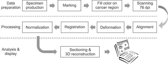
Figure 1 The overall processing diagram of the 3D distribution analysis of prostate cancer.
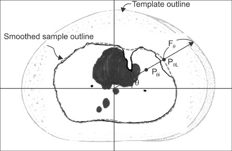
Figure 2 Template made from textbook and deformation procedure.
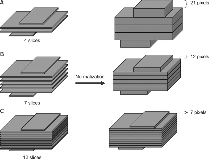
Figure 3 Normalization of samples which have different specimen sizes. Sample (A) has less slices for normal. So the thickness is increased to 21 pixels for each slice. (C) Sample has more slices for normal. So the thickness is reduced to 7 pixels for each slice.
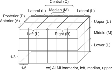
Figure 4 Definition of 3-dimensional distribution sections.
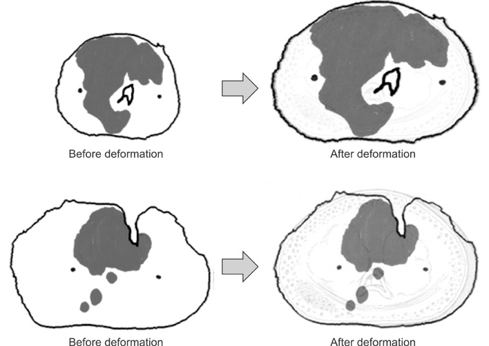
Figure 5 Deformation results.
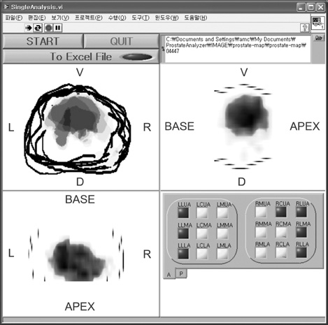
Figure 6 Three-dimensional distribution of prostate cancer using single sample analysis.
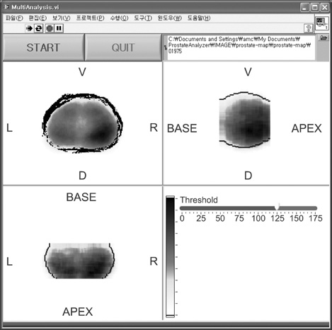
Figure 7 Three-dimensional distribution of prostate cancer using multi-sample analysis.
Reference
-
1. Korea Central Cancer Registry. Annual report of cancer statistics in Korean in 2008. 2011. 1st ed. Goyang: Korea Central Cancer Registry;24.2. True LD. Surgical pathology examination of the prostate gland. Practice survey by American society of clinical pathologists. Am J Clin Pathol. 1994. 102:572–579.
Article3. Zeng J, Bauer JJ, Mun SK. Modeling and mapping of prostate cancer. Comput Graph. 2000. 24:683–694.
Article4. Egevad L, Frimmel H, Norberg M, Mattson S, Carlbom I, Bengtsson E, Busch C. Three-dimensional computer reconstruction of prostate cancer from radical prostatectomy specimens: evaluation of the model by core biopsy simulation. Urology. 1999. 53:192–198.
Article5. Loch T, Chen ME. Computer simulation of prostate biopsies. Eur Urol Suppl. 2002. 1(6):47–51.
Article6. Mazzucchelli R, Santinelli A, Lopez-Beltran A, Scarpelli M, Montironi R. Evaluation of prognostic factors in radical prostatectomy specimens with cancer. Urol Int. 2002. 68:209–215.
Article7. Kang T, Song C, Song GH, Shin GH, Shin DI, Kim CS, Ahn H. The anatomic distribution and pathological characteristics of prostate cancer: a mapping analysis. Korean J Urol. 2006. 47:578–585.
Article8. Frimmel H, Egevad L, Bengtsson E, Busch C. Modeling prostate cancer distributions. Urology. 1999. 54:1028–1034.
Article9. Song C, Kim J, Chung H, Kim CS, Ro JY, Ahn HJ. Nomograms for the prediction of the pathological stage of the clinically localized prostate cancer in Korean men. Korean J Urol. 2003. 44:753–758.10. Lee SE, Kim DY, Kwak C. Interrelationship among age, prostate specific antigen and prostate volume in Korean men living at the metropolitan area. Korean J Urol. 1999. 40:1311–1317.11. Hodge KK, McNeal JE, Terris MK, Stamey TA. Random systematic versus directed ultrasound guided transrectal core biopsies of the prostate. J Urol. 1989. 142:71–74.
Article12. Vachani C. Understanding your pathology report: prostate cancer. cited at 2010 Dec 22. Available from: http://www.oncolink.org/types/article.cfm?c=16&s=57&ss=452&id=9597.13. Goshtasby A, Turner DA, Ackerman LV. Matching of tomographic slices for interpolation. IEEE Trans Med Imaging. 1992. 11:507–516.
Article14. Mazzucchelli R, Santinelli A, Lopez-Beltran A, Scarpelli M, Montironi R. Evaluation of prognostic factors in radical prostatectomy specimens with cancer. Urol Int. 2002. 68:209–215.
Article15. McNeal JE, Redwine EA, Freiha FS, Stamey TA. Zonal distribution of prostatic adenocarcinoma. Correlation with histologic pattern and direction of spread. Am J Surg Pathol. 1988. 12:897–906.16. Chen ME, Johnston DA, Tang K, Babaian RJ, Troncoso P. Detailed mapping of prostate carcinoma foci: biopsy strategy implications. Cancer. 2000. 89:1800–1809.
- Full Text Links
-
- Actions
-
Cited
- CITED
-
- Close
- Share
-
- Similar articles
-
- Screening for Prostatic Cancers in Korean
- Design & Development of 3D Medical Image Processing System using VTK
- Diagnostic Value for Early Detection of Prostate Cancer of the Digital Rectal Examination, Serum Prostate Antigen and Transrectal Ultrasonography
- The prostate specific antigen in detection of the prostate cancer
- The Anatomic Distribution and Pathological Characteristics of Prostate Cancer: A Mapping Analysis
