Formation of ghost images due to metal objects on the surface of the patient's face: A pictorial essay
- Affiliations
-
- 1Department of Oral Radiology, School of Dentistry, Pontifical Catholic University of Minas Gerais, Belo Horizonte, Brazil. manzi@pucminas.br
- 2Department of Oral Radiology, School of Dentistry, Federal University of Pará, Belém do Pará, Brazil.
- KMID: 2160160
- DOI: http://doi.org/10.5624/isd.2016.46.1.63
Abstract
- Panoramic radiographs are a relatively simple technique that is commonly used in all dental specialties. In panoramic radiographs, in addition to the formation of real images of metal objects, ghost images may also form, and these ghost images can hinder an accurate diagnosis and interfere with the accuracy of radiology reports. Dentists must understand the formation of these images in order to avoid making incorrect radiographic diagnoses. Therefore, the present study sought to present a study of the formation of panoramic radiograph ghost images caused by metal objects in the head and neck region of a dry skull, as well as to report a clinical case n order to warn dentists about ghost images and to raise awareness thereof. An understanding of the principles of the formation of ghost images in panoramic radiographs helps prevent incorrect diagnoses.
MeSH Terms
Figure
-
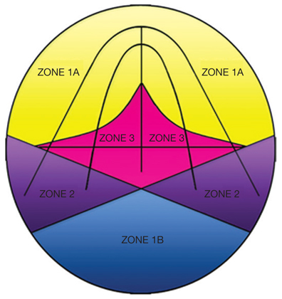
Fig. 1 The diagram shows zones 1A and 1B (formation of one image), zone 2 (formation of two images), and zone 3 (formation of three images).
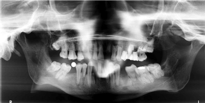
Fig. 2 A panoramic radiograph with a metal sphere attached in the region where the right mandibular first molar is missing (zone 1A) shows a single real image of the metal sphere.
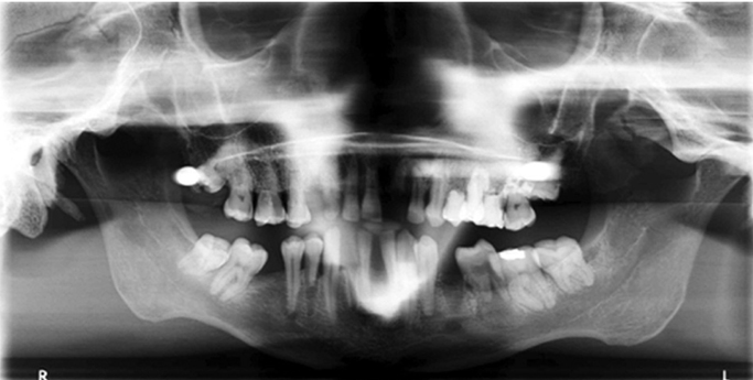
Fig. 3 A panoramic radiograph with a metal sphere attached in the region of the crown of the right maxillary third molar (zone 2) shows two images: one real ipsilateral image and one contralateral ghost image.
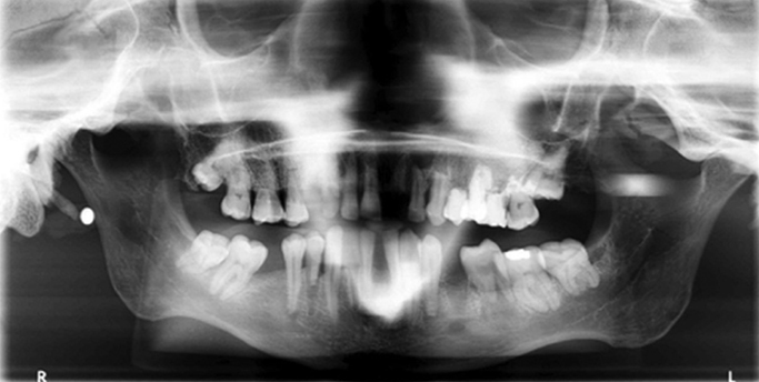
Fig. 4 A panoramic radiograph with a metal sphere attached in the region of the right earlobe (zone 2) shows two images: one real ipsilateral image and one distorted contralateral ghost image.
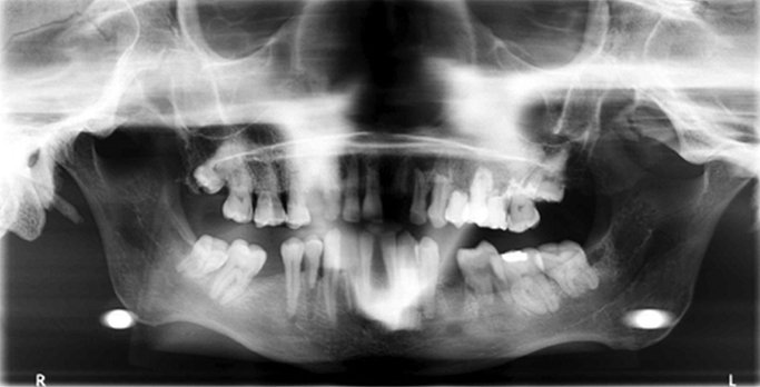
Fig. 5 A panoramic radiograph with a metal sphere attached in the region of the hyoid bone (zone 3) shows two real and bilateral images (mandibular angle region) and one ghost image in the center of the image receptor.
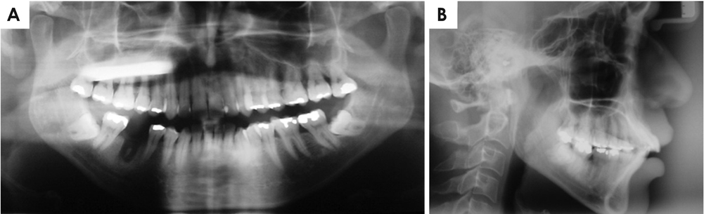
Fig. 6 A. A panoramic radiograph shows a radiopaque band overlapping the periapical region of the right maxillary molars, premolars, and canine. Additionally, a voluminous metal restoration is observed in the occlusal region of the left maxillary third molar. B. A lateral cephalometric radiograph shows that no other metal object is present in the head.
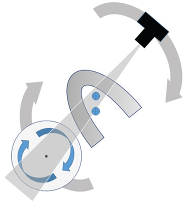
Fig. 7 This illustration shows the production of standard panoramic images. The image receptor and X-ray source rotate around the patient's head. During the exposure cycle, the center of rotation of the X-ray beam changes position.
Reference
-
1. American Dental Association (ADA). Food and Drug Administration (FDA). Council on Scientific Affairs [Internet]. Dental radiographic examinations; recommendations for patient selection and limiting radiation exposure. 2012. 1–27. Available from: http://www.ada.org/~/media/ADA/Member Center/FIles/Dental_Radiographic_Examinations_2012.ashx.2. Kaugars GE, Collett WK. Panoramic ghosts. Oral Surg Oral Med Oral Pathol. 1987; 63:103–108.
Article3. Pittayapat P, Willems G, Alqerban A, Coucke W, Ribeiro-Rotta RF, Souza PC, et al. Agreement between cone beam computed tomography images and panoramic radiographs for initial orthodontic evaluation. Oral Surg Oral Med Oral Pathol Oral Radiol. 2014; 117:111–119.
Article4. Perschbacher S. Interpretation of panoramic radiographs. Aust Dent J. 2012; 57:Suppl 1. 40–45.
Article5. Ladeira DB, Cruz AD, Almeida SM, Bóscolo FN. Influence of the intergonial distance on image distortion in panoramic radiographs. Dentomaxillofac Radiol. 2012; 41:417–421.
Article6. Catić A, Celebić A, Valentić-Peruzović M, Catović A, Jerolimov V, Muretić I. Evaluation of the precision of dimensional measurements of the mandible on panoramic radiographs. Oral Surg Oral Med Oral Pathol Oral Radiol Endod. 1998; 86:242–248.7. Valerio CS, Trindade AM, Mazzieiro ET, Amaral TP, Manzi FR. Use of digital panoramic radiography as an auxiliary means of low bone mineral density detection in post-menopausal women. Dentomaxillofac Radiol. 2013; 42:20120059.
Article8. Quintero JC, Trosien A, Hatcher D, Kapila S. Craniofacial imaging in orthodontics: historical perspective, current status, and future developments. Angle Orthod. 1999; 69:491–506.9. Kim YK, Park JY, Kim SG, Kim JS, Kim JD. Magnification rate of digital panoramic radiographs and its effectiveness for pre-operative assessment of dental implants. Dentomaxillofac Radiol. 2011; 40:76–83.
Article10. Monsour PA, Mendoza AR. Panoramic ghost images as an aid in the localization of soft tissue calcifications. Oral Surg Oral Med Oral Pathol. 1990; 69:748–756.
Article11. Reuter I, Ritter W, Kaeppler G. Triple images on panoramic radiographs. Dentomaxillofac Radiol. 1999; 28:316–319.
Article12. McDavid WD, Langlais RP, Welander U, Morris CR. Real, double, and ghost images in rotational panoramic radiography. Dentomaxillofac Radiol. 1983; 12:122–128.
Article13. White SC, Pharoah MJ. Oral radiology; principles and interpretation. 6rd ed. St. Louis: Mosby-Year Book Inc;2009.14. McDavid WD, Dove SB, Welander U, Tronje G. Dimensional reproduction in direct digital rotational panoramic radiography. Oral Surg Oral Med Oral Pathol. 1993; 75:523–527.
Article
- Full Text Links
-
- Actions
-
Cited
- CITED
-
- Close
- Share
-
- Similar articles
-
- An atypical case involving real, ghost, and pseudo-ghost images on a panoramic radiograph
- Pictorial, Essay : Sonography of the Stomach
- Multi-Detector CT Findings of Typical and Atypical Appendicitis: A Pictorial Essay
- Imaging Features of Inflammatory Breast Disorders: A Pictorial Essay
- Penile Incarceration with Metal and Plastic Objects
