Calcaneal Osteomyelitis due to Non-tuberculous Mycobacteria: A Case Report
- Affiliations
-
- 1Department of Rehabilitation Medicine, Bundang Jesaeng General Hospital, Seongnam, Korea. hsa12345@hanmail.net
- 2Department of Pathology, Bundang Jesaeng General Hospital, Seongnam, Korea.
- KMID: 2155181
- DOI: http://doi.org/10.5535/arm.2016.40.1.172
Abstract
- Osteomyelitis is a bone infection caused by bacteria or other germs. Gram-positive cocci are the most common etiological organisms of calcaneal osteomyelitis; whereas, non-tuberculous mycobacteria (NTM) are rarely documented. We reported a case of NTM calcaneal osteomyelitis in a 51-year-old female patient. She had been previously treated in many local clinics with multiple local steroid injection over 50 times and extracorporeal shock-wave therapy over 20 times with the impression of plantar fasciitis for 3 years prior. Diagnostic workup revealed a calcaneal osteomyelitis and polymerase chain reaction assay on bone aspirate specimens confirmed the diagnosis of non-tuberculous mycobacterial osteomyelitis. The patient had a partial calcanectomy with antitubercular therapy. Six months after surgery, a follow-up magnetic resonance imaging showed localized chronic osteomyelitis with abscess formation. We continued anti-tubercular therapy without operation. At 18-month follow-up after surgery and comprehensive rehabilitation therapy, she was ambulating normally and able to carry out her daily activities without any discomfort.
MeSH Terms
Figure
-
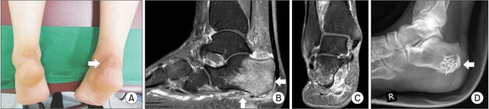
Fig. 1 (A) Mild swelling and color change over the right calcaneus. (B) Sagittal and (C) axial fat-suppressed T2-weighted magnetic resonance scan demonstrates cortical destruction at right medial calcaneal tuberosity with surrounding soft tissue inflammation and swelling over plantar aspect. (D) The operation site was packed with geneX bone graft substitute.
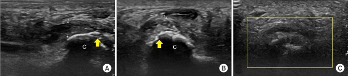
Fig. 2 (A) Longitudinal scan of right plantar heel. The plantar fascia was grossly thickened and measured 5.6 mm (distance between crosses) at the anteroinferior border of the calcaneus, 'c'. (B) Transverse scan of right plantar heel. Plantar aspect of right calcaneus showed cortical irregularity (yellow arrows) and hypoechoic swelling with edematous change in the surrounding soft tissues. (C) Color Doppler ultrasonography showed no vascularity in the right calcaneus.
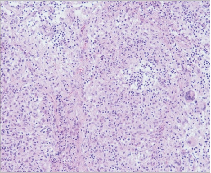
Fig. 3 Low power (200×) hematoxylin and eosin stained specimen showed diffuse ill-defined granulomatous inflammation with Langerhans giant cells that is highly suggestive of a mycobacterial infection.

Fig. 4 (A) Sagittal and (B) axial T2-weighted magnetic resonance scan of right foot. The image showed relative well-marginated osteolytic lesion with loculated fluid collection and adjacent thick peripheral rim enhancement of right calcaneus posterior portion, which is demonstrated as localized chronic stage osteomyelitis with abscess formation.
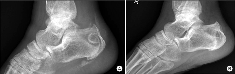
Fig. 5 (A) Lateral view of the foot showed predominantly lytic lesion involving the calcaneus. (B) At the 18-month follow-up, radiograph showed remineralization of bones with sclerosis of calcaneus.
Reference
-
1. Roxas M. Plantar fasciitis: diagnosis and therapeutic considerations. Altern Med Rev. 2005; 10:83–93. PMID: 15989378.2. Thomas JL, Christensen JC, Kravitz SR, Mendicino RW, Schuberth JM, Vanore JV. The diagnosis and treatment of heel pain: a clinical practice guideline-revision 2010. J Foot Ankle Surg. 2010; 49(3 Suppl):S1–S19. PMID: 20439021.
Article3. Elsayed S, Read R. Mycobacterium haemophilum osteomyelitis: case report and review of the literature. BMC Infect Dis. 2006; 6:70. PMID: 16606464.
Article4. Michelarakis J, Varouhaki C. Osteomyelitis of the calcaneus due to atypical Mycobacterium. Foot Ankle Surg. 2009; 15:106–108. PMID: 19410179.
Article5. Hwang JH, Koh WJ, Kim EJ, Kang EH, Suh GY, Chung MP, et al. Partial interferon-gamma receptor deficiency and non-tuberculous mycobacterial lung disease. Tuberculosis (Edinb). 2006; 86:382–385. PMID: 16682253.6. Healey K, Chen K. Plantar fasciitis: current diagnostic modalities and treatments. Clin Podiatr Med Surg. 2010; 27:369–380. PMID: 20691370.
Article7. Gidumal R, Evanski P. Calcaneal osteomyelitis following steroid injection: a case report. Foot Ankle. 1985; 6:44–46. PMID: 4043891.
Article8. Wronka KS, Sinha A. Calcaneal osteomyelitis following steroid injection for plantar fasciitis: a case report. Foot Ankle Spec. 2012; 5:253–255. PMID: 22732240.
- Full Text Links
-
- Actions
-
Cited
- CITED
-
- Close
- Share
-
- Similar articles
-
- Calcaneal Osteomyelitis Presenting as a Paradoxical Reaction during Treatment of Multidrug-Resistant Tuberculosis
- Tuberculous Osteomyelitis of the Tarsal Bone in an Infant: Case Report
- One Stage Management with Curettage and Bone Graft of Chronic Osteomyelitis Preceded by Surgical Treatment of Calcaneal Fracture: Report of Three Cases
- Vertebral Osteomyelitis caused by Mycobacterium abscessus in an Immunocompetent Patient
- Tuberculous Osteomyelitis on the Proximal Humerus after BCG vaccination: a case report
