J Korean Orthop Assoc.
2016 Feb;51(1):91-95. 10.4055/jkoa.2016.51.1.91.
Diffuse-Type Giant Cell Tumor Arising from a Pretibial Lesion: Extra-Articular Pigmented Villonodular Tenosynovitis
- Affiliations
-
- 1Department of Orthopaedic Surgery, Daejeon Sun General Hospital, Daejeon, Korea. mydangjang@naver.com
- KMID: 2154354
- DOI: http://doi.org/10.4055/jkoa.2016.51.1.91
Abstract
- Pigmented villonodular synovitis (PVNS) is a rare, benign, soft tissue neoplasm affecting the synovium of joints, classified as localized and diffused type. Localized type is more common, arising from synovium of joints, bursae, and tendon sheaths. Diffused type is relatively rare, frequently arising from an extra-articular lesion, and sometimes from an intramuscular or subcutaneous lesion. Although the cause of occurrence is not yet clear, recently it has been known as a benign neoplasm rather than an inflammatory or reactive process. We performed a total excision of the PVNS in a pretibial lesion and achieved a good result. We report on the case with a review of the literature.
MeSH Terms
Figure
-
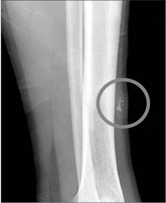
Figure 1 Lateral radiograph shows a radiopaque lesion (circle) in the pretibial lesion, the left.
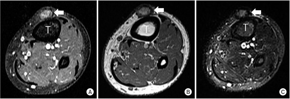
Figure 2 Magnetic resonance imaging findings. T: tibia, arrow: mass lesion. (A) Axial spin-echo T1-weighted image after contrast injection shows a focal hypointense area, findings that represent the blurring effect from hemosiderin. (B, C) Axial T1- and T2-weighted images show the localized intermediate signal intensity soft tissue mass at the same level.
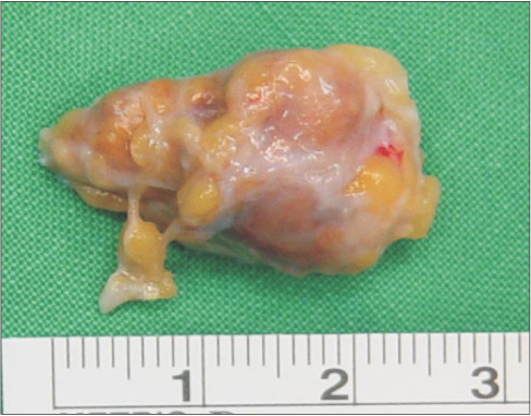
Figure 3 Photograph of the resected specimen shows the brownish appearance caused by hemosiderin.
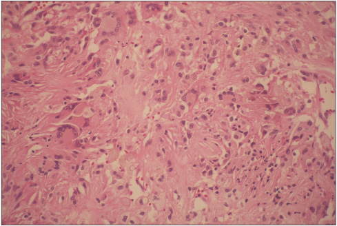
Figure 4 Microscopic finding. The picture shows multinucleated giant cells, hemosiderin deposits, and foam cells (H&E, ×400).
Reference
-
1. Brien EW, Sacoman DM, Mirra JM. Pigmented villonodular synovitis of the foot and ankle. Foot Ankle Int. 2004; 25:908–913.
Article2. Murphey MD, Rhee JH, Lewis RB, Fanburg-Smith JC, Flemming DJ, Walker EA. Pigmented villonodular synovitis: radiologic-pathologic correlation. Radiographics. 2008; 28:1493–1518.
Article3. Sanghvi DA, Purandare NC, Jambhekar NA, Agarwal MG, Agarwal A. Diffuse-type giant cell tumor of the subcutaneous thigh. Skeletal Radiol. 2007; 36:327–330.
Article4. Rochwerger A, Groulier P, Curvale G, Launay F. Pigmented villonodular synovitis of the foot and ankle: a report of eight cases. Foot Ankle Int. 1999; 20:587–590.
Article5. Saxena A, Perez H. Pigmented villonodular synovitis about the ankle: a review of the literature and presentation in 10 athletic patients. Foot Ankle Int. 2004; 25:819–826.
Article6. Riccio AI, Christoforetti J, Annunziata CC. Pigmented villonodular synovitis of the pes anserine bursa: case report. J Knee Surg. 2007; 20:44–47.7. Jelinek JS, Kransdorf MJ, Utz JA, et al. Imaging of pigmented villonodular synovitis with emphasis on MR imaging. AJR Am J Roentgenol. 1989; 152:337–342.
Article8. Darling JM, Goldring SR, Harada Y, Handel ML, Glowacki J, Gravallese EM. Multinucleated cells in pigmented villonodular synovitis and giant cell tumor of tendon sheath express features of osteoclasts. Am J Pathol. 1997; 150:1383–1393.9. Flandry F, Hughston JC. Pigmented villonodular synovitis. J Bone Joint Surg Am. 1987; 69:942–949.
Article10. Schwartz HS, Unni KK, Pritchard DJ. Pigmented villonodular synovitis. A retrospective review of affected large joints. Clin Orthop Relat Res. 1989; 247:243–255.
- Full Text Links
-
- Actions
-
Cited
- CITED
-
- Close
- Share
-
- Similar articles
-
- Diffuse Pigmented Villonodular Synovitis of the Hip Joint
- Nodular Pigmented Villonodular Synovitis of the Right Shoulder Joint: One Case Report
- A Case of Diffuse Pigmented Villonodular Synovitis of the Hip Joint
- Pigmented Villonodular Synovitis of the Temporomandibular Joint: a case report
- Localized Pigmented Villonodular Synovitis of the Knee with Concomitant Intra- and Extra- Articular Lesion: A Case Report
