Anatomical Variant of Atlas : Arcuate Foramen, Occpitalization of Atlas, and Defect of Posterior Arch of Atlas
- Affiliations
-
- 1Pohang SM Christianity Hospital, Pohang, Korea. hanibalkms@hanmail.net
- KMID: 2151154
- DOI: http://doi.org/10.3340/jkns.2015.58.6.528
Abstract
OBJECTIVE
We sought to examine anatomic variations of the atlas and the clinical significance of these variations.
METHODS
We retrospectively reviewed 1029 cervical 3-dimensional (3D) CT images. Cervical 3D CT was performed between November 2011 and August 2014. Arcuate foramina were classified as partial or complete and left and/or right. Occipitalization of the atlas was classified in accordance with criteria specified by Mudaliar et al. Posterior arch defects of the atlas were classified in accordance with criteria specified by Currarino et al.
RESULTS
One hundred and eight vertebrae (108/1029, 10.5%) showed an arcuate foramen. Bilateral arcuate foramina were present in 41 of these vertebrae and the remaining 67 arcuate foramina were unilateral (right 31, left 36). Right-side arcuate foramina were partial on 18 sides and complete on 54 sides. Left-side arcuate foramina were partial on 24 sides and complete on 53 sides. One case of atlas assimilation was found. Twelve patients (12/1029, 1.17%) had a defect of the atlantal posterior arch. Nine of these patients (9/1029, 0.87%) had a type A posterior arch defect. We also identified one type B, one type D, and one type E defect.
CONCLUSION
Preoperative diagnosis of occipitalization of the atlas and arcuate foramina using 3D CT is of paramount importance in avoiding neurovascular injury during surgery. It is important to be aware of posterior arch defects of the atlas because they may be misdiagnosed as a fracture.
Figure
-

Fig. 1 Classification of posterior arch defects of the atlas. Type A : failure of posterior midline fusion of the two hemiarches. Sometimes a small separate ossicle is seen within the gap, Type B : unilateral cleft, ranging from a small gap to a complete absence of the hemiarch and posterior tubercle, Type C : bilateral clefts of the lateral aspects of the arches with preservation of the most dorsal part of the arch, Type D : complete absence of the posterior arch with a persistent isolated tubercle. This anomaly is conceivably a more extensive form of bilateral clefts. The lateral parts of the posterior arch are absent except for the posterior tubercle, Type E : absence of the entire posterior arch including the tubercle. Reproduced with permission from Currarino G, Rollins N, Diehl JT : Congenital defects of the posterior arch of the atlas : a report of seven cases including an affected mother and son. AJNR Am J Neuroradiol 15 : 249-254, 19945).

Fig. 2 Arcuate foramen. A : Three-dimensional computed tomography of a 21-year-old man showing complete arcuate foramina on both sides (arrows). B : Three-dimensional computed tomography of a 54-year-old woman demonstrating a partial arcuate foramen on her right side (arrow).
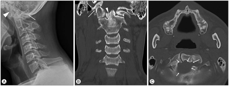
Fig. 3 Occipitalization of the atlas. A : Lateral cervical spine X-ray image of a 32-year-old man showing occipitalization of the atlas, C2–3 fusion (arrow : fusion between occiput and posterior arch of atlas, arrowhead : fusion between occiput and anterior arch of atlas). B : Coronal computed tomography demonstrating an abnormal odontoid process (arrowhead) and occipitalization of the atlas (arrow). C : Axial computed tomography showing an anomalous foramen magnum (arrows).

Fig. 4 Defect of the posterior arch of the atlas. A and B : Three-dimensional computed tomography of a 34-year-old man showing a Type A defect of the posterior arch of the atlas (A, arrow) and a defect of the anterior arch of the atlas (B, arrow, arrowhead : no defect in upper portion of anterior arch). C : Three-dimensional computed tomography of a 58-year-old woman showing a Type B defect (arrow). D and E : Three-dimensional computed tomography of a 41-year-old woman showing a Type D defect (arrow). F and G : Lateral cervical spine X-ray image (F, arrow : absence of the entire arch, including the tubercle) and three-dimensional computed tomography (G, arrow : absence of the entire arch, including the tubercle) of a 14-year-old girl showing a Type E defect.
Reference
-
1. Ahmad FU, Wang MY. Lateral mass of C1 fixation and ponticulus-posticus. World Neurosurg. 2014; 82:e145–e146. PMID: 24530457.
Article2. Al-Motabagani MA, Surendra M. Total occipitalization of the atlas. Anat Sci Int. 2006; 81:173–180. PMID: 16955668.
Article3. Bailey DK. The normal cervical spine in infants and children. Radiology. 1952; 59:712–719. PMID: 12994006.
Article4. Cakmak O, Gurdal E, Ekinci G, Yildiz E, Cavdar S. Arcuate foramen and its clinical significance. Saudi Med J. 2005; 26:1409–1413. PMID: 16155658.5. Currarino G, Rollins N, Diehl JT. Congenital defects of the posterior arch of the atlas : a report of seven cases including an affected mother and son. AJNR Am J Neuroradiol. 1994; 15:249–254. PMID: 8192068.6. Cushing KE, Ramesh V, Gardner-Medwin D, Todd NV, Gholkar A, Baxter P, et al. Tethering of the vertebral artery in the congenital arcuate foramen of the atlas vertebra : a possible cause of vertebral artery dissection in children. Dev Med Child Neurol. 2001; 43:491–496. PMID: 11463182.
Article7. Erbengi A, Oge HK. Congenital malformations of the craniovertebral junction : classification and surgical treatment. Acta Neurochir (Wien). 1994; 127:180–185. PMID: 7942200.
Article8. Garg A, Gaikwad SB, Gupta V, Mishra NK, Kale SS, Singh J. Bipartite atlas with os odontoideum : case report. Spine (Phila Pa 1976). 2004; 29:E35–E38. PMID: 14722424.9. Geipel P. [Studies on the fissure formation of the atlas and epistropheus. IV]. Zentralbl Allg Pathol. 1955; 94:19–84. PMID: 13282406.10. Gupta S, Goel A. Quantitative anatomy of the lateral masses of the atlas and axis vertebrae. Neurol India. 2000; 48:120–125. PMID: 10878774.11. Harrower G. Variations in the region of the foramen magnum. J Anat. 1923; 57(Pt 2):178–192. PMID: 17103966.12. Hong JT, Jang WY, Kim IS, Yang SH, Sung JH, Son BC, et al. Posterior C1 stabilization using superior lateral mass as an entry point in a case with vertebral artery anomaly : technical case report. Neurosurgery. 2011; 68(1 Suppl Operative):246–249. discussion 249PMID: 21206308.13. Huang MJ, Glaser JA. Complete arcuate foramen precluding C1 lateral mass screw fixation in a patient with rheumatoid arthritis : case report. Iowa Orthop J. 2003; 23:96–99. PMID: 14575258.14. Kassim NM, Latiff AA, Das S, Ghafar NA, Suhaimi FH, Othman F, et al. Atlanto-occipital fusion : an osteological study with clinical implications. Bratisl Lek Listy. 2010; 111:562–565. PMID: 21125803.15. Klimo P Jr, Blumenthal DT, Couldwell WT. Congenital partial aplasia of the posterior arch of the atlas causing myelopathy : case report and review of the literature. Spine (Phila Pa 1976). 2003; 28:E224–E228. PMID: 12811285.16. Koutsouraki E, Avdelidi E, Michmizos D, Kapsali SE, Costa V, Baloyannis S. Kimmerle's anomaly as a possible causative factor of chronic tension-type headaches and neurosensory hearing loss : case report and literature review. Int J Neurosci. 2010; 120:236–239. PMID: 20374094.
Article17. Lamberty BG, Zivanović S. The retro-articular vertebral artery ring of the atlas and its significance. Acta Anat (Basel). 1973; 85:113–122. PMID: 4197316.
Article18. Li S, Li W, Sun J. [Operative treatment for cervical vertigo caused by foramen arcuale]. Zhonghua Wai Ke Za Zhi. 1995; 33:137–139. PMID: 7555378.19. Limousin CA. Foramen arcuale and syndrome of Barre-Lieou. Its surgical treatment. Int Orthop. 1980; 4:19–23. PMID: 7399776.20. Manjunath KY. Posterior bridging of the atlas vertebra in south Indians. Indian J Med Sci. 2001; 55:488–490. PMID: 11887298.21. Menezes AH. Craniocervical developmental anatomy and its implications. Childs Nerv Syst. 2008; 24:1109–1122. PMID: 18401563.
Article22. Mitchell J. The incidence of the lateral bridge of the atlas vertebra. J Anat. 1998; 193(Pt 2):283–285. PMID: 9827643.
Article23. Mudaliar RP, Shetty S, Nanjundaiah K, Kumar JP, Kc J. An osteological study of occipitocervical synostosis : its embryological and clinical significance. J Clin Diagn Res. 2013; 7:1835–1837. PMID: 24179875.24. Paraskevas G, Papaziogas B, Tsonidis C, Kapetanos G. Gross morphology of the bridges over the vertebral artery groove on the atlas. Surg Radiol Anat. 2005; 27:129–136. PMID: 15800734.
Article25. Penning L. Functional pathology of cervical spine. ed 1. London: Excerpta medica foundation;1968. Vol. 4:p. 76.26. Piper JG, Traynelis VC. Congenital malformations of the craniovertebral junction. In : Dickman CA, Spetzler RF, Sonntag VKH, editors. Surgery of the craniovertebral junction. New York: Thieme Medical Publishers;1998. p. 123–149.27. Richardson EG, Boone SC, Reid RL. Intermittent quadriparesis associated with a congenital anomaly of the posterior arch of the atlas. Case report. J Bone Joint Surg Am. 1975; 57:853–854. PMID: 1158928.
Article28. Sagiuchi T, Tachibana S, Sato K, Shimizu S, Kobayashi I, Oka H, et al. Lhermitte sign during yawning associated with congenital partial aplasia of the posterior arch of the atlas. AJNR Am J Neuroradiol. 2006; 27:258–260. PMID: 16484387.29. Sakai K, Tsutsui T. Bow hunter's stroke associated with atlantooccipital assimilation--case report. Neurol Med Chir (Tokyo). 1999; 39:696–700. PMID: 10563123.
Article30. Sharma A, Gaikwad SB, Deol PS, Mishra NK, Kale SS. Partial aplasia of the posterior arch of the atlas with an isolated posterior arch remnant : findings in three cases. AJNR Am J Neuroradiol. 2000; 21:1167–1171. PMID: 10871035.31. Skrzat J, Mróz I, Jaworek JK, Walocha J. A case of occipitalization in the human skull. Folia Morphol (Warsz). 2010; 69:134–137. PMID: 21154282.32. Split W, Sawrasewicz-Rybak M. [Clinical symptoms and signs in Kimmerle anomaly]. Wiad Lek. 2002; 55:416–422. PMID: 12428570.33. Torreman M, Verhagen IT, Sluzewski M, Kok AJ, van Rooij WJ. Recurrent transient quadriparesis after minor cervical trauma associated with bilateral partial agenesis of the posterior arch of the atlas. Case report. J Neurosurg. 1996; 84:663–665. PMID: 8613860.
Article34. Travan L, Saccheri P, Sabbadini G, Crivellato E. Bilateral arcuate foramen associated with partial defect of the posterior arch of the atlas in a medieval skeleton : case report and review of the literature. Looking backward to go forward. Surg Radiol Anat. 2011; 33:495–500. PMID: 21153641.
Article35. Tubbs RS, Shoja MM, Shokouhi G, Farahani RM, Loukas M, Oakes WJ. Simultaneous lateral and posterior ponticles resulting in the formation of a vertebral artery tunnel of the atlas : case report and review of the literature. Folia Neuropathol. 2007; 45:43–46. PMID: 17357011.36. Wang S, Wang C, Liu Y, Yan M, Zhou H. Anomalous vertebral artery in craniovertebral junction with occipitalization of the atlas. Spine (Phila Pa 1976). 2009; 34:2838–2842. PMID: 20010391.
Article37. Wight S, Osborne N, Breen AC. Incidence of ponticulus posterior of the atlas in migraine and cervicogenic headache. J Manipulative Physiol Ther. 1999; 22:15–20. PMID: 10029944.
Article38. Wysocki J, Bubrowski M, Reymond J, Kwiatkowski J. Anatomical variants of the cervical vertebrae and the first thoracic vertebra in man. Folia Morphol (Warsz). 2003; 62:357–363. PMID: 14655117.
- Full Text Links
-
- Actions
-
Cited
- CITED
-
- Close
- Share
-
- Similar articles
-
- Cerebellar Ectopia Associated with Unilateral Agenesis of Posterior Arch of Atlas
- Congenital Anomaly of the Atlas Misdiagnosed as Posterior Arch Fracture of the Atlas and Atlantoaxial Subluxation
- Hypertrophic Posterior Arch of Atlas Causing Cervical Myelopathy
- Combined Congenital Anterior and Posterior Midline Cleft of the Atlas Associated with Asymptomatic Lateral Atlantoaxial Subluxation
- Stress Fracture of the Anterior Atlas Arch Following C1 Posterior Arch Resection for Cervical Myelopathy with Retro-Odontoid Pseudotumor
