Isolated Cerebellar Variant of Adrenoleukodystrophy with a de novo Adenosine Triphosphate-Binding Cassette D1 (ABCD1) Gene Mutation
- Affiliations
-
- 1Department of Pediatrics, Chungnam National University Hospital, Chungnam National University College of Medicine, Daejeon, Korea.
- 2Division of Pediatric Neurology, Department of Pediatrics, Severance Children's Hospital, Yonsei University College of Medicine, Seoul, Korea. hipo0207@yuhs.ac
- 3Department of Clinical Genetics, Yonsei University College of Medicine, Seoul, Korea.
- 4Department of Pediatrics, Gangnam Severance Hospital, Yonsei University College of Medicine, Seoul, Korea.
- 5Department of Neurology, Yonsei University College of Medicine, Seoul, Korea.
- KMID: 2130847
- DOI: http://doi.org/10.3349/ymj.2014.55.4.1157
Abstract
- X-linked adrenoleukodystrophy (X-ALD) shows a wide range of phenotypic expression, but clinical presentation as an isolated lesion of the cerebellar white matter and dentate nuclei has not been reported. We report an unusual presentation of X-ALD only with an isolated lesion of the cerebellar white matter and dentate nuclei. The proband, a 37-year-old man presented with bladder incontinence, slurred speech, dysmetria in all limbs, difficulties in balancing, and gait ataxia. Brain magnetic resonance imaging showed an isolated signal change of white matter around the dentate nucleus in cerebellum. With high level of very long chain fatty acid, gene study showed a de novo mutation in exon 1 at nucleotide position c.277_296dup20 (p.Ala100Cysfs*10) of the adenosine triphosphate-binding cassette D1 gene. It is advised to consider X-ALD as a differential diagnosis in patients with isolated cerebellar degeneration symptoms.
MeSH Terms
Figure
-
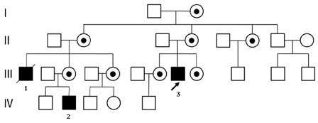
Fig. 1 Pedigree of the present family. The proband is indicated by an arrow. Affected males are designated by solid symbols and carrier females by circle with dot. Squares, male; circles, females.
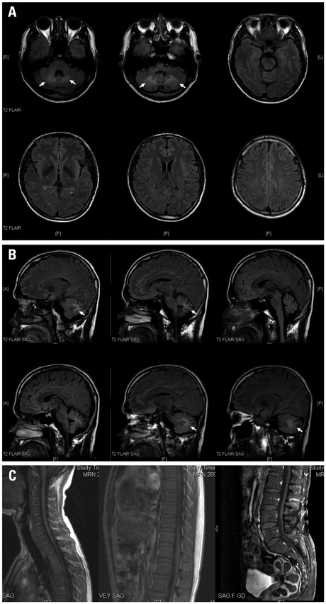
Fig. 2 (A) T2 magnetic resonance imaging (MRI) of the brain showed hypointense lesions in the dentate nuclei on axial planes. (B) T2-fluid-attenuated inversion recovery MRI of the brain showed hyperintense lesions in the dentate nuclei on sagittal planes. (C) No contrast enhancement was seen in T1-gadolium enhancement MRI of the spinal cord on sagittal plane.
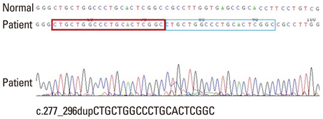
Fig. 3 Direct sequencing analysis of the proband with X-linked adrenoleukodystrophy demonstrated a novel duplication in exon 1 of adenosine triphosphate-binding cassette D1 gene, c.277_296dup20 (p.Ala100Cysfs*10). Box indicates position of the novel mutation.
Reference
-
1. Kemp S, Berger J, Aubourg P. X-linked adrenoleukodystrophy: clinical, metabolic, genetic and pathophysiological aspects. Biochim Biophys Acta. 2012; 1822:1465–1474.
Article2. Moser HW, Moser AB, Smith KD, Bergin A, Borel J, Shankroff J, et al. Adrenoleukodystrophy: phenotypic variability and implications for therapy. J Inherit Metab Dis. 1992; 15:645–664.3. Kusaka H, Imai T. Ataxic variant of adrenoleukodystrophy: MRI and CT findings. J Neurol. 1992; 239:307–310.
Article4. Dunne E, Hyman NM, Huson SM, Németh AH. A novel point mutation in X-linked adrenoleukodystrophy presenting as a spinocerebellar degeneration. Ann Neurol. 1999; 45:652–655.
Article5. Li JY, Hsu CC, Tsai CR. Spinocerebellar variant of adrenoleukodystrophy with a novel ABCD1 gene mutation. J Neurol Sci. 2010; 290:163–165.
Article6. Nakazato T, Sato T, Nakamura T, Tsuji S, Narabayashi H. Adrenoleukodystrophy presenting as spinocerebellar degeneration. Eur Neurol. 1989; 29:229–234.
Article7. Takada K, Onoda K, Takahashi K, Nakamura H, Taketomi T. An adult case of adrenoleukodystrophy with features of olivo-ponto-cerebellar atrophy: I. Clinical and pathological studies. Jpn J Exp Med. 1987; 57:53–58.8. Tan EK, Lim SH, Chan LL, Wong MC, Tan KP. X-linked adrenoleukodystrophy: spinocerebellar variant. Clin Neurol Neurosurg. 1999; 101:137–140.
Article9. Vianello M, Manara R, Betterle C, Tavolato B, Mariniello B, Giometto B. X-linked adrenoleukodystrophy with olivopontocerebellar atrophy. Eur J Neurol. 2005; 12:912–914.
Article10. Suzuki Y, Takemoto Y, Shimozawa N, Imanaka T, Kato S, Furuya H, et al. Natural history of X-linked adrenoleukodystrophy in Japan. Brain Dev. 2005; 27:353–357.
Article11. Kurihara K, Kim H, Tamura N, Iwasaki S, Hamaguchi K. [A case of adrenoleukodystrophy presenting large lesion of the cerebellar white matter and dentate nuclei on brain CT and MRI]. Rinsho Shinkeigaku. 1991; 31:72–78.12. Miyai I, Fujimura H, Umekage T, Watase K, Ueno S, Yorifuji S, et al. Magnetic resonance imaging in adrenoleukodystrophy presenting as spinocerebellar degeneration. J Neurol Neurosurg Psychiatry. 1990; 53:623–624.
Article13. Waragai M, Takaya Y, Hayashi M, Shibata N, Kobayashi M. MRI of adrenoleukodystrophy involving predominantly the cerebellum and brain stem. Neuroradiology. 1996; 38:788–791.
Article14. Mishra S, Modi M, Das CP, Prabhakar S. Adrenoleukodystrophy manifesting as spinocerebellar degeneration. Neurol India. 2006; 54:195–196.15. Loes DJ, Fatemi A, Melhem ER, Gupte N, Bezman L, Moser HW, et al. Analysis of MRI patterns aids prediction of progression in X-linked adrenoleukodystrophy. Neurology. 2003; 61:369–374.
Article16. Elenein RA, Naik S, Kim S, Punia V, Jin K. Teaching neuroimages: cerebral adrenoleukodystrophy: a rare adult form. Neurology. 2013; 80:e69–e70.
Article17. Poll-The BT, Engelen M. Peroxisomal leukoencephalopathy. Semin Neurol. 2012; 32:42–50.
Article
- Full Text Links
-
- Actions
-
Cited
- CITED
-
- Close
- Share
-
- Similar articles
-
- Novel ABCD1 Gene Mutation in a Korean Patient with X-Linked Adrenoleukodystrophy Presenting with Addison's Disease
- An Incidentally Identified Sporadic Case with Adrenoleukodystrophy with the ABCD1 Mutation
- Adrenomyeloneuropathy with cerebral involvement due to a novel frameshift variant in ABCD1 gene
- Cerebral Adrenomyeloneuropathy with Trp77-Leu82del Mutation in ABCD1 Gene
- Transient neonatal diabetes mellitus caused by a de novo ABCC8 gene mutation
