Imaging Sci Dent.
2015 Mar;45(1):15-22. 10.5624/isd.2015.45.1.15.
Reproducibility of the sella turcica landmark in three dimensions using a sella turcica-specific reference system
- Affiliations
-
- 1OIC, OMFS-IMPATH Research Group, Department of Imaging and Pathology, Faculty of Medicine, University of Leuven and Oral and Maxillofacial Surgery, University Hospitals Leuven, Leuven, Belgium. p.pittayapat@gmail.com
- 2Department of Radiology, Faculty of Dentistry, Chulalongkorn University, Bangkok, Thailand.
- 3Service de Chirurgie Orthopedique et Traumatologique, Centre Hospitalier Regional d'Orleans, Orleans Cedex 2, France.
- 4Department of Oral Diagnosis, Division of Oral Radiology, Piracicaba Dental School, University of Campinas, Piracicaba, Sao Paulo, Brazil.
- 5Orthodontics, Department of Oral Health Sciences, KU Leuven and Dentistry, University Hospitals Leuven, University of Leuven, Leuven, Belgium.
- 6Department of Oral and Maxillofacial Surgery, Cliniques Universitaires Saint Luc, Universite Catholique de Louvain, Brussels, Belgium.
- KMID: 2116787
- DOI: http://doi.org/10.5624/isd.2015.45.1.15
Abstract
- PURPOSE
This study was performed to assess the reproducibility of identifying the sella turcica landmark in a threedimensional (3D) model by using a new sella-specific landmark reference system.
MATERIALS AND METHODS
Thirty-two cone-beam computed tomographic scans (3D Accuitomo(R) 170, J. Morita, Kyoto, Japan) were retrospectively collected. The 3D data were exported into the Digital Imaging and Communications in Medicine standard and then imported into the Maxilim(R) software (Medicim NV, Sint-Niklaas, Belgium) to create 3D surface models. Five observers identified four osseous landmarks in order to create the reference frame and then identified two sella landmarks. The x, y, and z coordinates of each landmark were exported. The observations were repeated after four weeks. Statistical analysis was performed using the multiple paired t-test with Bonferroni correction (intraobserver precision: p<0.005, interobserver precision: p<0.0011).
RESULTS
The intraobserver mean precision of all landmarks was <1 mm. Significant differences were found when comparing the intraobserver precision of each observer (p<0.005). For the sella landmarks, the intraobserver mean precision ranged from 0.43+/-0.34 mm to 0.51+/-0.46 mm. The intraobserver reproducibility was generally good. The overall interobserver mean precision was <1 mm. Significant differences between each pair of observers for all anatomical landmarks were found (p<0.0011). The interobserver reproducibility of sella landmarks was good, with >50% precision in locating the landmark within 1 mm.
CONCLUSION
A newly developed reference system offers high precision and reproducibility for sella turcica identification in a 3D model without being based on two-dimensional images derived from 3D data.
Keyword
MeSH Terms
Figure
-
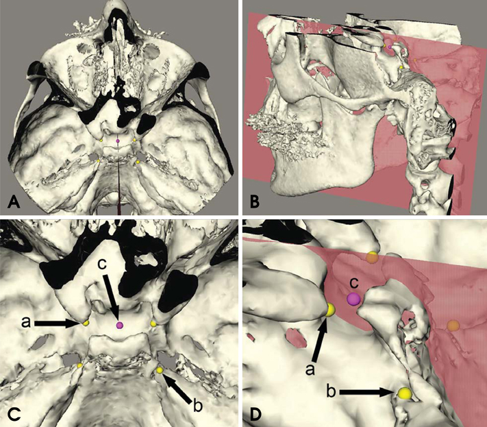
Fig. 1 (A) and (B) show a threedimensional model of one patient in a top and oblique overview, respectively. In this figure, a reference system is created by locating four landmarks (the right and left anterior clinoid process [ACP] and the right and left apex of the petrous part of the temporal bone right [APT]). (C) is a close-up of the top view presented in (A), showing the landmarks that form the reference system: (a) right and left ACP, (b) right and left APT, and (c) one of the sella landmarks. (D) is a close-up of the oblique view, showing landmarks (a), (b), and (c). The sella landmark (c) was located on one of the vertical planes created from the reference system in the Maxilim® software.
Reference
-
1. Alkofide EA. Sella turcica morphology and dimensions in cleft subjects. Cleft Palate Craniofac J. 2008; 45:647–653.
Article2. Axelsson S, Storhaug K, Kjaer I. Post-natal size and morphology of the sella turcica. Longitudinal cephalometric standards for Norwegians between 6 and 21 years of age. Eur J Orthod. 2004; 26:597–604.
Article3. Brock-Jacobsen MT, Pallisgaard C, Kjaer I. The morphology of the sella turcica in monozygotic twins. Twin Res Hum Genet. 2009; 12:598–604.
Article4. Kjær I. Sella turcica morphology and the pituitary gland - a new contribution to craniofacial diagnostics based on histology and neuroradiology. Eur J Orthod. 2015; 37:28–36.5. Teal JS. Radiology of the adult sella turcica. Bull Los Angeles Neurol Soc. 1977; 42:111–174.6. Alkofide EA. The shape and size of the sella turcica in skeletal Class I, Class II, and Class III Saudi subjects. Eur J Orthod. 2007; 29:457–463.
Article7. Andredaki M, Koumantanou A, Dorotheou D, Halazonetis DJ. A cephalometric morphometric study of the sella turcica. Eur J Orthod. 2007; 29:449–456.
Article8. Hofrath H. Bedeutung der Röntgenfern und Abstands Aufnahme für die Diagnostik der Kieferanomalien. Fortschr Orthod. 1931; 1:231–258.9. Broadbent BH. A new x-ray technique and its application to orthodontia. Angle Orthod. 1931; 1:45–66.10. Pauwels R, Beinsberger J, Collaert B, Theodorakou C, Rogers J, Walker A, et al. Effective dose range for dental cone beam computed tomography scanners. Eur J Radiol. 2012; 81:267–271.
Article11. Mah JK, Huang JC, Choo H. Practical applications of conebeam computed tomography in orthodontics. J Am Dent Assoc. 2010; 141:7S–13S.
Article12. Kau CH, Richmond S, Palomo JM, Hans MG. Three-dimensional cone beam computerized tomography in orthodontics. J Orthod. 2005; 32:282–293.13. van Vlijmen OJ, Kuijpers MA, Bergé SJ, Schols JG, Maal TJ, Breuning H, et al. Evidence supporting the use of cone-beam computed tomography in orthodontics. J Am Dent Assoc. 2012; 143:241–252.
Article14. Swennen GR, Schutyser F, Barth EL, De Groeve P, De Mey A. A new method of 3-D cephalometry Part I: the anatomic Cartesian 3-D reference system. J Craniofac Surg. 2006; 17:314–325.15. Olszewski R, Cosnard G, Macq B, Mahy P, Reychler H. 3D CT-based cephalometric analysis: 3D cephalometric theoretical concept and software. Neuroradiology. 2006; 48:853–862.
Article16. Periago DR, Scarfe WC, Moshiri M, Scheetz JP, Silveira AM, Farman AG. Linear accuracy and reliability of cone beam CT derived 3-dimensional images constructed using an orthodontic volumetric rendering program. Angle Orthod. 2008; 78:387–395.
Article17. Brown AA, Scarfe WC, Scheetz JP, Silveira AM, Farman AG. Linear accuracy of cone beam CT derived 3D images. Angle Orthod. 2009; 79:150–157.
Article18. Olszewski R, Tanesy O, Cosnard G, Zech F, Reychler H. Reproducibility of osseous landmarks used for computed tomography based three-dimensional cephalometric analyses. J Craniomaxillofac Surg. 2010; 38:214–221.
Article19. Olszewski R, Frison L, Wisniewski M, Denis JM, Vynckier S, Cosnard G, et al. Reproducibility of three-dimensional cephalometric landmarks in cone-beam and low-dose computed tomography. Clin Oral Investig. 2013; 17:285–292.
Article20. Pittayapat P, Limchaichana-Bolstad N, Willems G, Jacobs R. Three-dimensional cephalometric analysis in orthodontics: a systematic review. Orthod Craniofac Res. 2014; 17:69–91.
Article21. Schlicher W, Nielsen I, Huang JC, Maki K, Hatcher DC, Miller AJ. Consistency and precision of landmark identification in three-dimensional cone beam computed tomography scans. Eur J Orthod. 2012; 34:263–275.
Article22. Chien PC, Parks ET, Eraso F, Hartsfield JK, Roberts WE, Ofner S. Comparison of reliability in anatomical landmark identification using two-dimensional digital cephalometrics and three-dimensional cone beam computed tomography in vivo. Dentomaxillofac Radiol. 2009; 38:262–273.23. de Oliveira AE, Cevidanes LH, Phillips C, Motta A, Burke B, Tyndall D. Observer reliability of three-dimensional cephalometric landmark identification on cone-beam computerized tomography. Oral Surg Oral Med Oral Pathol Oral Radiol Endod. 2009; 107:256–265.
Article24. Meyer-Marcotty P, Reuther T, Stellzig-Eisenhauer A. Bridging of the sella turcica in skeletal Class III subjects. Eur J Orthod. 2010; 32:148–153.
Article25. Cederberg RA, Benson BW, Nunn M, English JD. Calcification of the interclinoid and petroclinoid ligaments of sella turcica: a radiographic study of the prevalence. Orthod Craniofac Res. 2003; 6:227–232.
Article26. Bergland RM, Ray BS, Torack RM. Anatomical variations in the pituitary gland and adjacent structures in 225 human autopsy cases. J Neurosurg. 1968; 28:93–99.
Article27. Durão AR, Pittayapat P, Rockenbach MI, Olszewski R, Ng S, Ferreira AP, et al. Validity of 2D lateral cephalometry in orthodontics: a systematic review. Prog Orthod. 2013; 14:31.
Article28. Muramatsu A, Nawa H, Kimura M. Reproducibility of maxillofacial anatomic landmarks on 3-dimensional computed tomographic images determined with the 95% confidence ellipse method. Angle Orthod. 2008; 78:396–402.
Article29. Hassan B, Nijkamp P, Verheij H, Tairie J, Vink C, van der Stelt P, et al. Precision of identifying cephalometric landmarks with cone beam computed tomography in vivo. Eur J Orthod. 2013; 35:38–44.
Article
- Full Text Links
-
- Actions
-
Cited
- CITED
-
- Close
- Share
-
- Similar articles
-
- The evaluation of sella turcica on the shape and volume in class III patients
- Assessment of the Relationship between Sella Turcica Morphology and Delayed Dental Age
- The study of shape and size of normal sella turcica in cephalometric radiographs
- Ossifying fibroma of the sella turcica
- Empty sella syndrome
