J Korean Soc Spine Surg.
2001 Jun;8(2):172-175. 10.4184/jkss.2001.8.2.172.
Insufficiency Fracture of the Sacrum: A Case Report
- Affiliations
-
- 1Department of Orthopaedic Surgery, Pohang St. Mary's Hospital, Korea. TotalHip@unitel.co.kr
- KMID: 2097758
- DOI: http://doi.org/10.4184/jkss.2001.8.2.172
Abstract
- Sacral insufficiency fractures are unexpected causes of inguinal, lower back and buttock pain in elderly women with osteoporosis who have sustained unknown or only minimal trauma. Differential and radiological diagnoses of these fractures are often difficult. Bone scan remains the standard diagnostic tool, but computed tomography or magnetic resonance image may be required to differentiate insufficiency fracture from other diseases such as malignant bone lesion. The fracture usually extends vertically in the sacral ala, parallel to the sacroiliac joints. This distribution suggests that such fractures could be partially caused by weight-bearing transmitted through the spine. We report the treatment of insufficiency sacral fracture in one osteopenic patient who has been confirmed by computed tomography and treated with conservative method that convinced by follow up computed tomography.
MeSH Terms
Figure
-

Fig. 1. Anteroposterior pelvic radiograph showing sclerosis in the left ala is otherwise unremarkable.
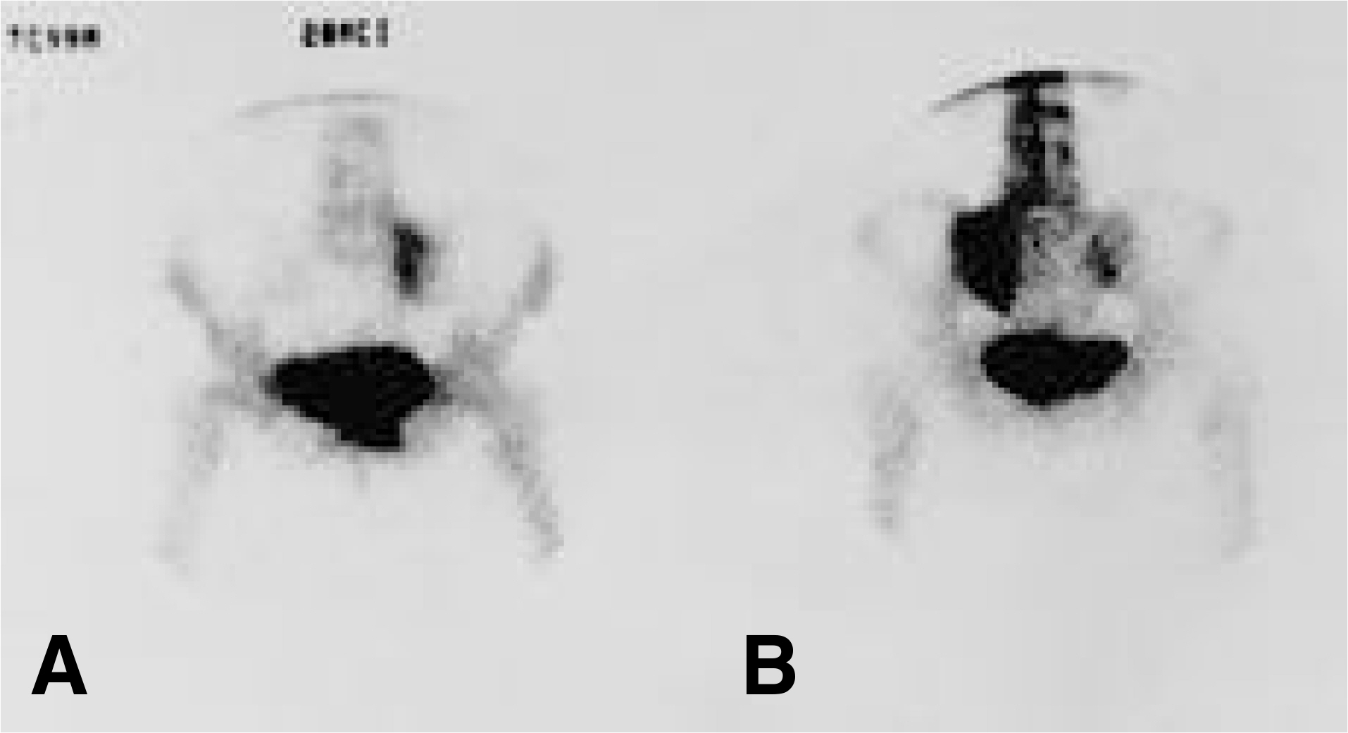
Fig. 2. Technetium 99m bone scan revealing increased uptake in the left sacral ala. Fig. A. supine position, B. prone position
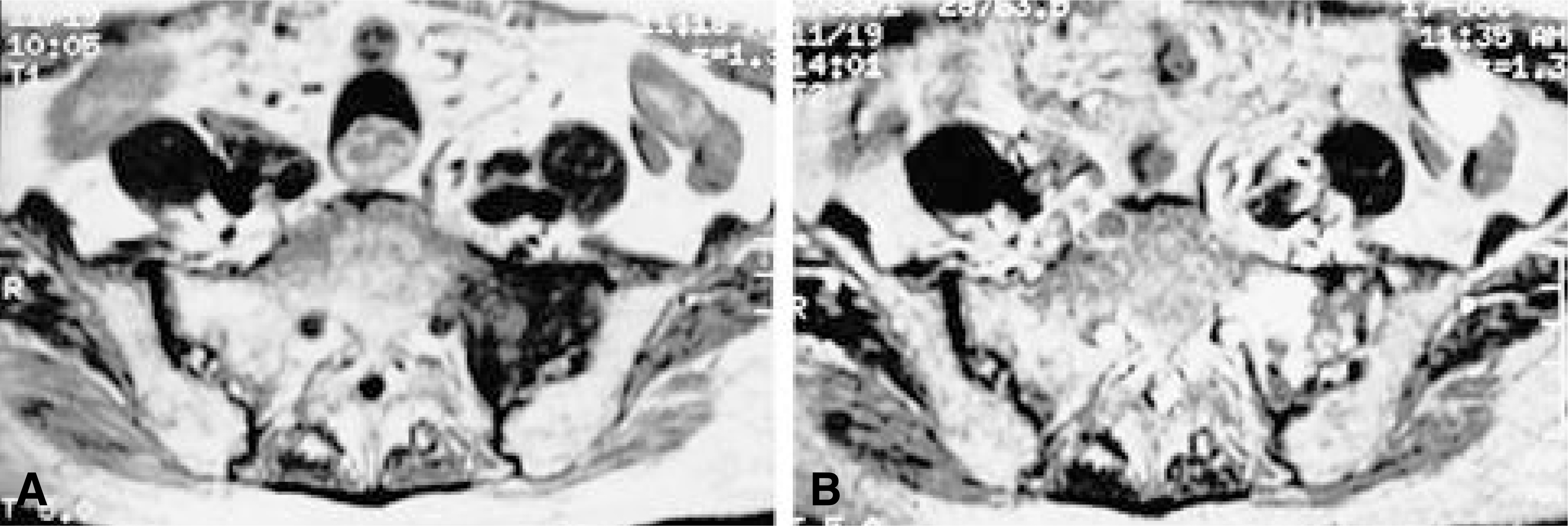
Fig. 3. T1 and T2-weighted axial images of the sacrum, demonstrating a lesion in the left sacral ala which shows low signal intensity on the T1 image (A) and high signal intensity on the T2 image (B).

Fig. 4. Computed tomographic section of the sacrum, demonstrating the left alar fracture parallel to the sacroiliac joint and broad band of sclerosis.

Fig. 5. 7 months followup computed tomography scan con-firms a healing left sacral ala fracture.
Reference
-
1). Cooper KL, Beabout JW, Swee RG. Insufficiency fractures of the sacrum. Radiology. 156:15–20. 1985.
Article2). Grangier C, Garcia J, Howarth NR, May M, Rossier P. Role of MRI in the diagnosis of insufficiency fractures of the sacrum and acetabular roof. Skeletal Radiol. 26:517–524. 1997.
Article3). Hauge MD, Cooper KL, Litin SC. I nsu fficien cy fractures of the pelvis that simulate metastatic disease. Mayo Clin Proc. 63:807–812. 1988.4). Jean LL, Bruno D, Eric T, Francis B, Francois B. Sacral insufficiency fractures presenting as acute low-back pain. Spine. 18:2502–2506. 1993.
Article5). Jung JH, Jun JB, Shim SC, et al. Insufficiency fractures of sacrum and pubic rami in rhematoid arthritis. J Korean Rheum Assoc. 5:287–292. 1998.6). Kim YS, Jo SS. The significance of bone scan in fatigue fractures. J of Korean Orthop Surgery. 19:189–196. 1984.7). Marcel W, Paul H, Heini G. Insufficiency fractures of the sacrum. Spine. 18:2507–2512. 1993.
Article8). Newhouse KE, Elkhoury GY, Buckwalter JA. Occult sacral fractures on osteopenic patients. J Bone Joint Surg. 74-A:1472–1477. 1992.9). Renner JB. Pelvic insufficiency fractures. Arthritis Rheum. 33:426–430. 1990.
Article10). Sterling GW, John LT, Michael RB, Howard MP. Sacral insufficiency fractures in rheumatoid arthritis. Spine. 19:2117–2121. 1994.
Article
- Full Text Links
-
- Actions
-
Cited
- CITED
-
- Close
- Share
-
- Similar articles
-
- Insufficiency Fracture of the Sacrum: Report of two cases
- Usefulness of Kyphoplasty in Sacral Insufficiency Fracture: A Case Report
- MR Findings of Sacral Insufficiency Fractures in Osteoporotic Patients: Two Cases Report
- Sacral Insufficiency Fracture, Usually Overlooked Cause of Lumbosacral Pain
- Dual Plate Fixation for Displaced Transverse Fracture of the Lower Sacrum
