Positioning errors and quality assessment in panoramic radiography
- Affiliations
-
- 1Department of Oral Medicine and Radiology, ITS Centre for Dental Studies and Research, Ghaziabad, India. drmanudhillon@yahoo.co.in
- 2Department of Oral Medicine and Radiology, Teerthanker Mahavir Dental College, Moradabad, India.
- 3Department of Oral Medicine and Radiology, Kothiwal Dental College and Research Centre, Moradabad, India.
- 4Department of Pedodontics and Preventive Dentistry, IDST Dental College and Research Centre, Modinagar, India.
- KMID: 1806785
- DOI: http://doi.org/10.5624/isd.2012.42.4.207
Abstract
- PURPOSE
This study was performed to determine the relative frequency of positioning errors, to identify those errors directly responsible for diagnostically inadequate images, and to assess the quality of panoramic radiographs in a sample of records collected from a dental college.
MATERIALS AND METHODS
This study consisted of 1,782 panoramic radiographs obtained from the Department of Oral and Maxillofacial Radiology. The positioning errors of the radiographs were assessed and categorized into nine groups: the chin tipped high, chin tipped low, a slumped position, the patient positioned forward, the patient positioned backward, failure to position the tongue against the palate, patient movement during exposure, the head tilted, and the head turned to one side. The quality of the radiographs was further judged as being 'excellent', 'diagnostically acceptable', or 'unacceptable'.
RESULTS
Out of 1,782 radiographs, 196 (11%) were error free and 1,586 (89%) were present with positioning errors. The most common error observed was the failure to position the tongue against the palate (55.7%) and the least commonly experienced error was patient movement during exposure (1.6%). Only 11% of the radiographs were excellent, 64.1% were diagnostically acceptable, and 24.9% were unacceptable.
CONCLUSION
The positioning errors found on panoramic radiographs were relatively common in our study. The quality of panoramic radiographs could be improved by careful attention to patient positioning.
Keyword
Figure
-
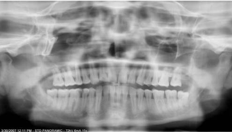
Fig. 1 The patient's chin is too high, causing a flat occlusal plane, splayed condyles, and loss of sharpness of the maxillary incisors.

Fig. 2 The patient's chin is too low. The occlusal plane is "smiling" and the apices of the mandibular incisors are fuzzy.

Fig. 3 The cervical spine is slumped, appearing as a pyramid shaped opacity, centred at the lower half of the film.
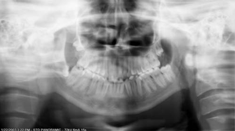
Fig. 4 The central incisors are in front of the bite groove, causing them to appear thin and fuzzy. The cervical spine is in the focal zone, causing it to be superimposed on the mandible.
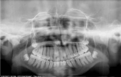
Fig. 5 The anterior teeth are positioned behind the bite groove causing them to appear wider than normal.

Fig. 6 Panoramic radiograph shows the patient failure to position the tongue against the palate leading to a dark shadow over the maxillary teeth between the palate and dorsum of the tongue.
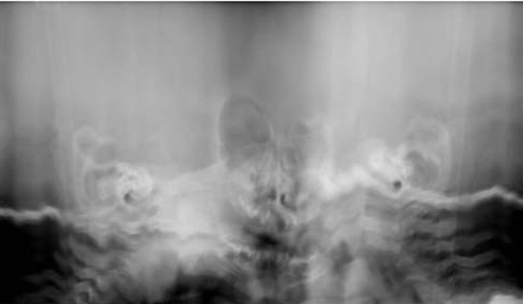
Fig. 7 Radiograph shows image distortion due to patient movement during exposure.
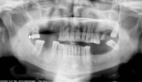
Fig. 8 The head is tilted to one side, causing one condyle to appear higher than the other and the inferior border of the mandible is slanting.

Fig. 9 The head is turned to one side, causing an asymmetry of the condyles, and wider teeth and ramus on one side than the other.
Cited by 2 articles
-
Common positioning errors in panoramic radiography: A review
Rafael Henrique Nunes Rondon, Yamba Carla Lara Pereira, Glauce Crivelaro do Nascimento
Imaging Sci Dent. 2014;44(1):1-6. doi: 10.5624/isd.2014.44.1.1.A new bite block for panoramic radiographs of anterior edentulous patients: A technical report
Jong-Woong Park, Khanthaly Symkhampha, Kyung-Hoe Huh, Won-Jin Yi, Min-Suk Heo, Sam-Sun Lee, Soon-Chul Choi
Imaging Sci Dent. 2015;45(2):117-122. doi: 10.5624/isd.2015.45.2.117.
Reference
-
1. Horner K. Review article: radiation protection in dental radiology. Br J Radiol. 1994. 67:1041–1049.2. Brezden NA, Brooks SL. Evaluation of panoramic dental radiographs taken in private practice. Oral Surg Oral Med Oral Pathol. 1987. 63:617–621.
Article3. Eliasson S, Lavstedt S, Wouters F, Ostlin L. Quality of intraoral radiographs sent by private dental practitioners for therapy evaluation by the Social Insurance Office. Swed Dent J. 1990. 14:81–89.4. Akesson L, Håkansson J, Rohlin M, Zöger B. An evaluation of image quality for the assessment of the marginal bone level in panoramic radiography. A comparison of radiographs from different dental clinics. Swed Dent J. 1993. 17:9–21.5. Svenson B, Eriksson T, Kronström M, Palmqvist S. Image quality of intraoral radiographs used by general practitioners in prosthodontic treatment planning. Dentomaxillofac Radiol. 1994. 23:46–48.
Article6. Svenson B, Eriksson T, Kronström M, Palmqvist S. Quality of intraoral radiographs used for prosthodontic treatment planning by general dentists in the public dental health service. Swed Dent J. 1995. 19:47–54.7. Rushton VE, Horner K, Worthington HV. The quality of panoramic radiographs in a sample of general dental practices. Br Dent J. 1999. 186:630–633.
Article8. Helminen SE, Vehkalahti M, Wolf J, Murtomaa H. Quality evaluation of young adults' radiographs in Finnish public oral health service. J Dent. 2000. 28:549–555.
Article9. Langland OE, Sippy FH, Morris CR, Langlais RP. Principles and practice of panoramic radiology. 1992. 2nd ed. Philadelphia: WB Saunders.10. White SC, Pharaoh MJ. Oral Radiology: principles and interpretation. 2004. 5th ed. Philadelphia: CV Mosby;200–217.11. Pitts NB, Kidd EA. Some of the factors to be considered in the prescription and timing of bitewing radiography in the diagnosis and management of dental caries. J Dent. 1992. 20:74–84.
Article12. Royal College of Radiologists. National Radiological Protection Board. Guidelines on radiology standards for primary dental care. 1994. London: NRPB;30.13. Frederiksen NL, Benson BW, Sokolowski TW. Effective dose and risk assessment from film tomography used for dental implant diagnostics. Dentomaxillofac Radiol. 1994. 23:123–127.
Article14. Nixon PP, Thorogood J, Holloway J, Smith NJ. An audit of film reject and repeat rates in a department of dental radiology. Br J Radiol. 1995. 68:1304–1307.
Article15. Choi BR, Choi DH, Huh KH, Yi WJ, Heo MS, Choi SC, et al. Clinical image quality evaluation for panoramic radiography in Korean dental clinics. Imaging Sci Dent. 2012. 42:183–190.
Article16. McDavid WD, Langlais RP, Welander U, Morris CR. Real, double, and ghost images in rotational panoramic radiography. Dentomaxillofac Radiol. 1983. 12:122–128.
Article17. Rushton VE, Horner K. The use of panoramic radiology in dental practice. J Dent. 1996. 24:185–201.
Article18. Cohen J. A coefficient of agreement for nominal scales. Educ Psychol Meas. 1960. 20:37–46.
Article19. Altman DG. Practical statistics for medical research. 1990. London: Chapman and Hall.20. Akarslan ZZ, Erten H, Güngör K, Celik I. Common errors on panoramic radiographs taken in a dental school. J Contemp Dent Pract. 2003. 4:24–34.
Article21. Rushton VE, Horner K, Worthington HV. Aspects of panoramic radiography in general dental practice. Br Dent J. 1999. 186:342–344.
Article
- Full Text Links
-
- Actions
-
Cited
- CITED
-
- Close
- Share
-
- Similar articles
-
- Common positioning errors in panoramic radiography: A review
- Positioning errors and quality assessment in panoramic radiography
- A new bite block for panoramic radiographs of anterior edentulous patients: A technical report
- Clinical image quality evaluation for panoramic radiography in Korean dental clinics
- Assessment of panoramic radiography as a national oral examination tool: review of the literature
