Evaluation of linear measurements of implant sites based on head orientation during acquisition: An ex vivo study using cone-beam computed tomography
- Affiliations
-
- 1Section of Oral and Maxillofacial Radiology, Department of Oral Diagnostic Sciences, University of Connecticut School of Dental Medicine, Farmington, CT, USA. Tadinada@uchc.edu
- 2Section of Prosthodontics, Department of Reconstructive Sciences, University of Connecticut School of Dental Medicine, Farmington, CT, USA.
- KMID: 2054208
- DOI: http://doi.org/10.5624/isd.2015.45.2.73
Abstract
- PURPOSE
This study evaluated the effect of various head orientations during cone-beam computed tomography (CBCT) image acquisition on linear measurements of potential implant sites.
MATERIALS AND METHODS
Six dry human skulls with a total of 28 implant sites were evaluated for seven different head orientations. The scans were acquired using a Hitachi CB-MercuRay CBCT machine. The scanned volumes were reconstructed. Horizontal and vertical measurements were made and were compared to measurements made after simulating the head position to corrected head angulations. Data was analyzed using a two-way ANOVA test.
RESULTS
Statistical analysis revealed a significant interaction between the mean errors in vertical measurements with a marked difference observed at the extension head position (P<0.05). Statistical analysis failed to yield any significant interaction between the mean errors in horizontal measurements at various head positions.
CONCLUSION
Head orientation could significantly affect the vertical measurements in CBCT scans. The main head position influencing the measurements is extension.
Keyword
MeSH Terms
Figure
-
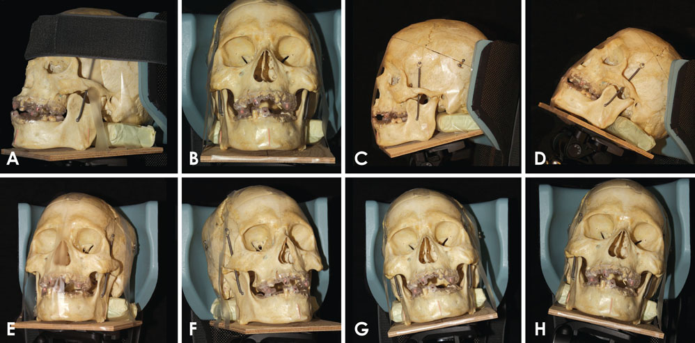
Fig. 1 Various head positions. A and B. Centric: The three tripod angles marked at zero degrees; it demonstrates zero x, y, and z axes. The occlusal plane and the Frankfort's horizontal plane is parallel to the floor. This position is considered to be the gold standard position. C. Flexion: The skull is tilted downward anteriorly by 20 degrees. D. Extension: The skull is tilted upward and backward by 20 degrees. E. Right flexion: The skull is moved 20 degrees laterally, away from the midline towards the right side. F. Left flexion: The skull is moved 20 degrees laterally, away from the midline towards the left side. G. Right lateral: The skull is directed 15 degrees towards the right shoulder. H. Left lateral: The skull is directed 15 degrees towards the left shoulder.
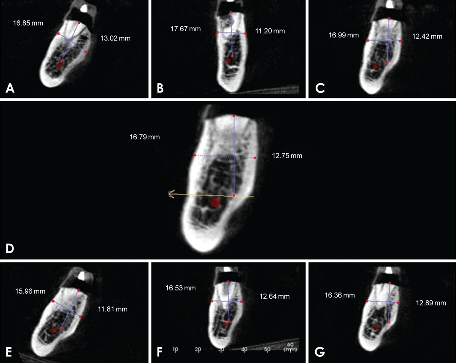
Fig. 2 Vertical and horizontal measurements of the alveolar ridge at a molar site at various head positions. A. Extension, B. Flexion, C. Right, D. Centric, E. Left, F. Right flexion, G. Left flexion.
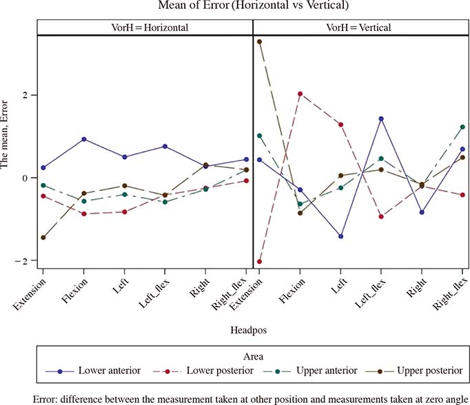
Fig. 3 Mean error of vertical and horizontal measurements for each head position at different implant sites.
Cited by 3 articles
-
The impact of reorienting cone-beam computed tomographic images in varied head positions on the coordinates of anatomical landmarks
Jae Hun Kim, Ho-Gul Jeong, Jae Joon Hwang, Jung-Hee Lee, Sang-Sun Han
Imaging Sci Dent. 2016;46(2):133-139. doi: 10.5624/isd.2016.46.2.133.Intraobserver and interobserver reproducibility in linear measurements on axial images obtained by cone-beam computed tomography
Nathália Cristine da Silva, Maurício Barriviera, José Luiz Cintra Junqueira, Francine Kühl Panzarella, Ricardo Raitz
Imaging Sci Dent. 2017;47(1):11-15. doi: 10.5624/isd.2017.47.1.11.Effect of slice inclination and object position within the field of view on the measurement accuracy of potential implant sites on cone-beam computed tomography
Bardia Vadiati Saberi, Negar Khosravifard, Alireza Nourzadeh
Imaging Sci Dent. 2020;50(1):37-43. doi: 10.5624/isd.2020.50.1.37.
Reference
-
1. Benavides E, Rios HF, Ganz SD, An CH, Resnik R, Reardon GT, et al. Use of cone beam computed tomography in implant dentistry: the International Congress of Oral Implantologists consensus report. Implant Dent. 2012; 21:78–86.2. Das KP, Jahangiri L, Katz RV. The first-choice standard of care for an edentulous mandible: a Delphi method survey of academic prosthodontists in the United States. J Am Dent Assoc. 2012; 143:881–889.3. Yim JH, Ryu DM, Lee BS, Kwon YD. Analysis of digitalized panorama and cone beam computed tomographic image distortion for the diagnosis of dental implant surgery. J Craniofac Surg. 2011; 22:669–673.
Article4. El-Beialy AR, Fayed MS, El-Bialy AM, Mostafa YA. Accuracy and reliability of cone-beam computed tomography measurements: influence of head orientation. Am J Orthod Dentofacial Orthop. 2011; 140:157–165.
Article5. Raes F, Renckens L, Aps J, Cosyn J, De Bruyn H. Reliability of circumferential bone level assessment around single implants in healed ridges and extraction sockets using cone beam CT. Clin Implant Dent Relat Res. 2013; 15:661–672.
Article6. Pertl L, Gashi-Cenkoglu B, Reichmann J, Jakse N, Pertl C. Preoperative assessment of the mandibular canal in implant surgery: comparison of rotational panoramic radiography (OPG), computed tomography (CT) and cone beam computed tomography (CBCT) for preoperative assessment in implant surgery. Eur J Oral Implantol. 2013; 6:73–80.7. Ganguly R, Ruprecht A, Vincent S, Hellstein J, Timmons S, Qian F. Accuracy of linear measurement in the Galileos cone beam computed tomography under simulated clinical conditions. Dentomaxillofac Radiol. 2011; 40:299–305.
Article8. Tsutsumi K, Chikui T, Okamura K, Yoshiura K. Accuracy of linear measurement and the measurement limits of thin objects with cone beam computed tomography: effects of measurement directions and of phantom locations in the fields of view. Int J Oral Maxillofac Implants. 2011; 26:91–100.9. Tarazona B, Llamas JM, Cibrian R, Gandia JL, Paredes V. A comparison between dental measurements taken from CBCT models and those taken from a digital method. Eur J Orthod. 2013; 35:1–6.
Article10. Leung CC, Palomo L, Griffith R, Hans MG. Accuracy and reliability of cone-beam computed tomography for measuring alveolar bone height and detecting bony dehiscences and fenestrations. Am J Orthod Dentofacial Orthop. 2010; 137:S109–S119.
Article11. Swennen GR, Schutyser F. Three-dimensional cephalometry: spiral multi-slice vs cone-beam computed tomography. Am J Orthod Dentofacial Orthop. 2006; 130:410–416.
Article12. Loubele M, Maes F, Schutyser F, Marchal G, Jacobs R, Suetens P. Assessment of bone segmentation quality of cone-beam CT versus multislice spiral CT: a pilot study. Oral Surg Oral Med Oral Pathol Oral Radiol Endod. 2006; 102:225–234.
Article13. Hassan B, van der Stelt P, Sanderink G. Accuracy of three-dimensional measurements obtained from cone beam computed tomography surface-rendered images for cephalometric analysis: influence of patient scanning position. Eur J Orthod. 2009; 31:129–134.
Article14. Araki K, Maki K, Seki K, Sakamaki K, Harata Y, Sakaino R, et al. Characteristics of a newly developed dentomaxillofacial X-ray cone beam CT scanner (CB MercuRay): system configuration and physical properties. Dentomaxillofac Radiol. 2004; 33:51–59.
Article15. Stratemann SA, Huang JC, Maki K, Miller AJ, Hatcher DC. Comparison of cone beam computed tomography imaging with physical measures. Dentomaxillofac Radiol. 2008; 37:80–93.
Article16. Moreira CR, Sales MA, Lopes PM, Cavalcanti MG. Assessment of linear and angular measurements on three-dimensional cone-beam computed tomographic images. Oral Surg Oral Med Oral Pathol Oral Radiol Endod. 2009; 108:430–436.
Article17. Frongia G, Piancino MG, Bracco P. Cone-beam computed tomography: accuracy of three-dimensional cephalometry analysis and influence of patient scanning position. J Craniofac Surg. 2012; 23:1038–1043.18. Sheikhi M, Ghorbanizadeh S, Abdinian M, Goroohi H, Badrian H. Accuracy of linear measurements of galileos cone beam computed tomography in normal and different head positions. Int J Dent. 2012; 2012:214954.
Article19. Visconti MA, Verner FS, Assis NM, Devito KL. Influence of maxillomandibular positioning in cone beam computed tomography for implant planning. Int J Oral Maxillofac Surg. 2013; 42:880–886.
Article
- Full Text Links
-
- Actions
-
Cited
- CITED
-
- Close
- Share
-
- Similar articles
-
- Diagnostic efficacy of a modified low-dose acquisition protocol for the preoperative evaluation of mini-implant sites
- Commentary on "Reliability of two different presurgical preparation methods for implant dentistry based on panoramic radiography and cone-beam computed tomography in cadavers"
- Reply on "Reliability of two different presurgical preparation methods for implant dentistry based on panoramic radiography and cone-beam computed tomography in cadavers"
- Comparison of cone-beam computed tomography cephalometric measurements using a midsagittal projection and conventional two-dimensional cephalometric measurements
- Does the metal artifact reduction algorithm activation mode influence the magnitude of artifacts in CBCT images?
