Qualitative changes in fetal trabecular meshwork fibers at the human iridocorneal angle
- Affiliations
-
- 1Division of Ophthalmology, Iwamizawa Municipal Hospital, Iwamizawa, Japan. fmhosaka@gmail.com
- 2Department of Anatomy and Embryology II, Faculty of Medicine, Complutense University, Madrid, Spain.
- 3Department of Anatomy, Akita University School of Medicine, Akita, Japan.
- 4Division of Internal Medicine, Iwamizawa Kojin-kai Hospital, Iwamizawa, Japan.
- 5Department of Anatomy, Sapporo Medical University, Sapporo, Japan.
- 6Department of Ophthalmology, Sapporo Medical University, Sapporo, Japan.
- KMID: 2046757
- DOI: http://doi.org/10.5115/acb.2013.46.1.49
Abstract
- We examined a series of changes that occur in the trabecular meshwork fibers of human eyes during fetal development at 12-30 weeks of gestation. At 12 and 15 weeks, the uveal meshwork was stained black with silver impregnation (indicating the predominance of collagen types III and IV) in the endomysium of the ciliary muscle. At 20 weeks, in combination with Schlemm's canal, a dense fibrous tissue mass corresponding to the trabecular meshwork anlage appeared and was colored black. The anlage was continuous with the corneal endothelium rather than with the ciliary muscle. Until 25 weeks, the trabecular meshwork was identifiable as fragmented fiber bundles that stained red-black, suggesting a mixture of collagen types I, III, and IV. At 30 weeks, half of the ciliary muscle fibers were inserted into the scleral spur and not into the meshwork. Therefore, any contribution of ciliary muscle contraction to the differentiation of the trabecular meshwork would appear to be limited. We hypothesize that an uneven distribution of mechanical stresses in the area of the cornea-sclera junction causes a tear thereby creating Schlemm's canal and is accompanied by a change in the collagen fiber types comprising the meshwork.
MeSH Terms
Figure
-
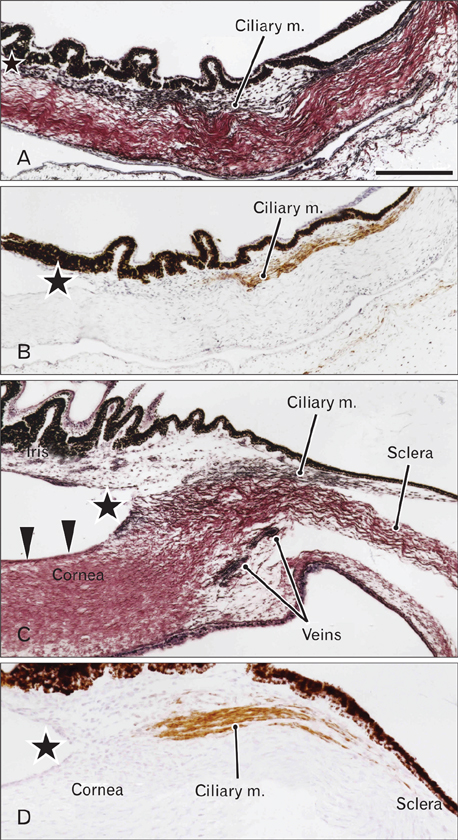
Fig. 1 Early stage of iridocorneal angle development. Horizontal sections. (A, C) Silver impregnation and (B, D) smooth muscle actin immunohistochemistry. Panels (A) and (B) display sections from a 12-week fetus, while (C) and (D) show sections from a 15-week fetus. At these stages, the iridocorneal angle (star) is distant from the ciliary muscle. Smooth muscle is immunopositive (B, D) and is accompanied by types III and IV collagen fibers (black fibers in A and C). In the cornea and sclera, type I collagen fibers appear red with silver staining. At 15 weeks, small spaces appear in the loose mesenchymal tissue at the angle. The corneal endothelium (arrowheads in C) is well preserved in this specimen. Scale bar in (A)=0.2 mm (A-D).
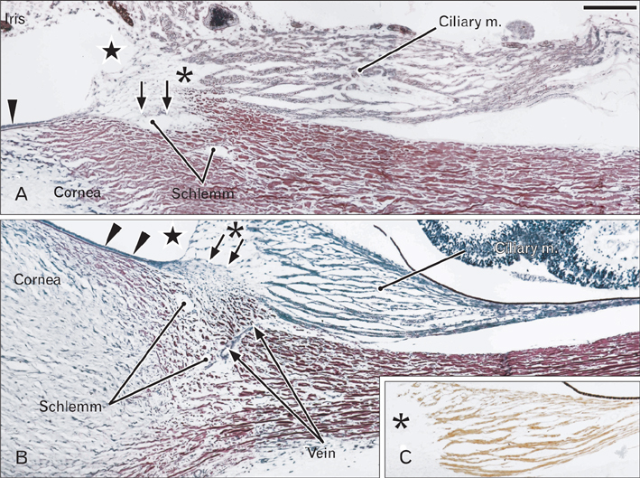
Fig. 2 Iridocorneal angle without a definite trabecular meshwork at 20 weeks. Horizontal sections. Silver impregnation (A, B) and smooth muscle actin immunohistochemistry (C). Panel (A) displays a specimen different from (B) and (C). Panel (C) shows a section taken from near (B). Schlemm's and collector canal (Schlemm) is evident in both specimens. On the corneal side of the ciliary muscle, a loose mesenchymal tissue, or the uveal meshwork, is evident (asterisk in A and B), but parts of the tissue form a relatively dense fibrous mass at the future trabecular meshwork (a perspective anlage; arrows in A and B). In (B), a thin vein passes through the margin of the sclera. The corneal endothelium is well preserved in these specimens (arrowheads in A and B). Scale bar in (A)=0.2 mm (A-C).
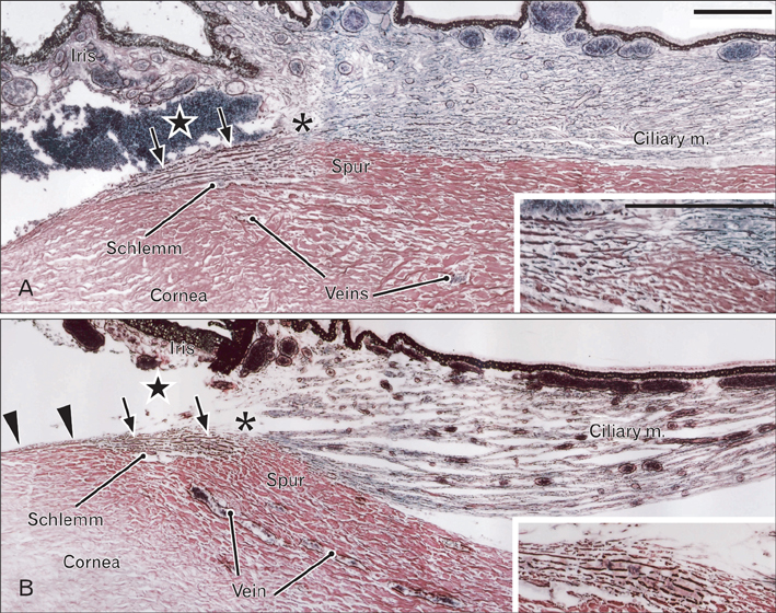
Fig. 3 Iridocorneal angle with a definite trabecular meshwork. Horizontal sections. Silver impregnation. Panel (A; panel B) displays a 25-week fetus (a 30-week fetus). In both specimens, the trabecular meshwork is evident (arrows) and is composed of red-black fiber bundles. Schlemm's canal (Schlemm) is adjacent to the meshwork. Insert at the lower right-hand angle is a higher magnification view of the meshwork. The brown fibers have collagen fiber types that differ from the red fibers in the cornea and sclera, as well as from the black fibers in the ciliary muscle. The sclera spur (spur) is evident in both specimens. The connection between the ciliary muscle and trabecular meshwork appears to be very weak because of loose interposing tissue (asterisk). A black-colored mass in the anterior chamber in (A) is blood, possibly due to injury during abortion. The corneal endothelium is well preserved in (B, arrowheads), and is continuous with the trabecular meshwork. Scale bars in (A)=0.2 mm (A, B).
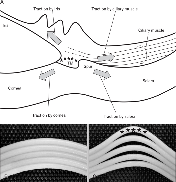
Fig. 4 Diagrams showing hypothetical induction of trabecular meshwork differentiation. Panel (A) displays four candidate generators of mechanical induction for early differentiation of the trabecular meshwork (TM): 1) growth of the iris, 2) contraction of the ciliary muscle, 3) thickening of the cornea, and 4) expansion of the sclera due to increased intraocular pressure. Panel (B) exhibits the lamellar configuration of the cornea and sclera in the early stage, while panel (C) shows lamellar distension and interlamellar dispersion due to sharing stress at the cornea-sclera junction. In (C), stars indicate the site at which the anlage of the TM appears. First, the sharing stress forms Schlemm's canal as a split, subsequently inducing the TM anlage to cover the tissue loss. Second, composite fibers of the TM anlage show a change in collagen types (becoming a mixture of types I, III, and IV), becoming fasciculated to form thick bundles and woven into a lattice-like meshwork.
Reference
-
1. McMenamin PG. Human fetal iridocorneal angle: a light and scanning electron microscopic study. Br J Ophthalmol. 1989. 73:871–879.2. McMenamin PG. A morphological study of the inner surface of the anterior chamber angle in pre and postnatal human eyes. Curr Eye Res. 1989. 8:727–739.3. McMenamin PG. A quantitative study of the prenatal development of the aqueous outflow system in the human eye. Exp Eye Res. 1991. 53:507–517.4. VanderWyst SS, Perkumas KM, Read AT, Overby DR, Stamer WD. Structural basement membrane components and corresponding integrins in Schlemm's canal endothelia. Mol Vis. 2011. 17:199–209.5. Kupfer C. A note on the development of the anterior chamber angle. Invest Ophthalmol. 1969. 8:69–74.6. Kupfer C, Ross K. The development of outflow facility in human eyes. Invest Ophthalmol. 1971. 10:513–517.7. Anderson DR. The development of the trabecular meshwork and its abnormality in primary infantile glaucoma. Trans Am Ophthalmol Soc. 1981. 79:458–485.8. Hansson HA, Jerndal T. Scanning electron microscopic studies on the development of the iridocorneal angle in human eyes. Invest Ophthalmol. 1971. 10:252–265.9. Ruano-Gil D, Costa-Vila J, Barastegui C. Arrangement of the sclerocorneal trabecular system in human fetuses. Acta Anat (Basel). 1986. 127:233–236.10. Tripathi RC. Davson H, Graham LT, editors. Comparative physiology and anatomy of the aqueous outflow pathway in the eye. The Eye. 1974. New York: Academic Press;163–336.11. Hogan MJ, Alvarado JA, Weddell JE. Histology of the human eye: an atlas and textbook. 1971. Philadelphia: W. B. Saunders.12. Meghpara B, Li X, Nakamura H, Khan A, Bejjani BA, Lin S, Edward DP. Human anterior chamber angle development without cell death or macrophage involvement. Mol Vis. 2008. 14:2492–2498.13. Marshall GE, Konstas AG, Lee WR. Collagens in ocular tissues. Br J Ophthalmol. 1993. 77:515–524.14. Abe S, Rhee SK, Osonoi M, Nakamura T, Cho BH, Murakami G, Ide Y. Expression of intermediate filaments at muscle insertions in human fetuses. J Anat. 2010. 217:167–173.15. Osanai H, Abe S, Rodríguez-Vázquez J, Verdugo-López S, Murakami G, Ohguro H. Human orbital muscle: a new point of view from the fetal development of extraocular connective tissues. Invest Ophthalmol Vis Sci. 2011. 52:1501–1506.16. Osanai H, Rodríguez-Vázquez JF, Abe H, Murakami G, Ohguro H, Fujimiya M. Fetal check ligament connected between the conjunctiva and the medial and lateral recti. Invest Ophthalmol Vis Sci. 2011. 52:7175–7179.17. Speakman JS. Drainage channels in the trabecular wall of Schlemm's canal. Br J Ophthalmol. 1960. 44:513–523.18. Lillie RD, Tracy RE, Pizzolato P, Donaldson PT, Reynolds C. Differential staining of collagen types in paraffin sections: a color change in degraded forms. Virchows Arch A Pathol Anat Histol. 1980. 386:153–159.19. Satoh T, Takeda R, Oikawa H, Satodate R. Immunohistochemical and structural characteristics of the reticular framework of the white pulp and marginal zone in the human spleen. Anat Rec. 1997. 249:486–494.20. Yamanouchi M. An electron microscopic study of the human iris vessels with special reference to the vascular changes on aging, using PAM-stain technique. Nihon Ganka Gakkai Zasshi. 1969. 73:767–784.21. Duance VC, Stephens HR, Dunn M, Bailey AJ, Dubowitz V. A role for collagen in the pathogenesis of muscular dystrophy? Nature. 1980. 284:470–472.22. Sevel D, Isaacs R. A re-evaluation of corneal development. Trans Am Ophthalmol Soc. 1988. 86:178–207.23. Rehnberg M, Ammitzböll T, Tengroth B. Collagen distribution in the lamina cribrosa and the trabecular meshwork of the human eye. Br J Ophthalmol. 1987. 71:886–892.24. Bystrom B, Virtanen I, Rousselle P, Gullberg D, Pedrosa-Domellof F. Distribution of laminins in the developing human eye. Invest Ophthalmol Vis Sci. 2006. 47:777–785.25. Ishii T. On the light microscopic fine structure of reticular fibers in the lymph node (a contribution to the understanding of the nature of reticular fibers). Verh Anat Ges. 1967. 62:487–494.26. Lutjen-Drecoll E, Futa R, Rohen JW. Ultrahistochemical studies on tangential sections of the trabecular meshwork in normal and glaucomatous eyes. Invest Ophthalmol Vis Sci. 1981. 21:563–573.27. Tamura Y, Konomi H, Sawada H, Takashima S, Nakajima A. Tissue distribution of type VIII collagen in human adult and fetal eyes. Invest Ophthalmol Vis Sci. 1991. 32:2636–2644.28. Grierson I, Rahi AH. Microfilaments in the cells of the human trabecular meshwork. Br J Ophthalmol. 1979. 63:3–8.29. Tumminia SJ, Mitton KP, Arora J, Zelenka P, Epstein DL, Russell P. Mechanical stretch alters the actin cytoskeletal network and signal transduction in human trabecular meshwork cells. Invest Ophthalmol Vis Sci. 1998. 39:1361–1371.30. Keller KE, Kelley MJ, Acott TS. Extracellular matrix gene alternative splicing by trabecular meshwork cells in response to mechanical stretching. Invest Ophthalmol Vis Sci. 2007. 48:1164–1172.31. Smelser GK, Ozanics V. The development of the trabecular meshwork in primate eyes. Am J Ophthalmol. 1971. 71(1 Pt 2):366–385.32. Johnstone MA. The aqueous outflow system as a mechanical pump: evidence from examination of tissue and aqueous movement in human and non-human primates. J Glaucoma. 2004. 13:421–438.33. Hamanaka T, Bill A, Ichinohasama R, Ishida T. Aspects of the development of Schlemm's canal. Exp Eye Res. 1992. 55:479–488.34. Smith RS, Zabaleta A, Savinova OV, John SW. The mouse anterior chamber angle and trabecular meshwork develop without cell death. BMC Dev Biol. 2001. 1:3.
- Full Text Links
-
- Actions
-
Cited
- CITED
-
- Close
- Share
-
- Similar articles
-
- Morphologic Changes of Anterior Chamber Angle and Trabecular Meshwork According to Embryonic Age in Human Fetal Eyes
- Age-related Changes of the Cellularity and Acid Mucopolysaccharides in the Trabecular Meshwork of the Normal Korean
- Change of Intraocular Pressure in Trabecular Meshwork Rupture Associated with Traumatic Hyphema
- Distribution of proteoglycans in the human trabecular meshwork: histochemical electron microscopic findings with cuprolinic blue
- The Effect of Accumulation of Mutant Myocilins in Human Trabecular Meshwork Cells
