Extramedullary Plasmacytoma from Bronchus Mimicking Lung Cancer: A Case Report
- Affiliations
-
- 1Department of Radiology, Yonsei University Wonju College of Medicine, Wonju Severance Christian Hospital, Wonju, Korea. andrew0668@hanmail.net
- 2Department of Pathology, Yonsei University Wonju College of Medicine, Wonju Severance Christian Hospital, Wonju, Korea.
- KMID: 2041934
- DOI: http://doi.org/10.3348/jksr.2014.70.4.251
Abstract
- Extramedullary plasmacytoma originating from the bronchus is a very rare condition, and the radiological diagnostic criteria for this disease are not well established due to its rarity. It often appears as a tumor with smooth margins and very rarely invades the surrounding structures. A computed tomography scan and a positron emission tomography/computed tomography scan were performed on a 71-year-old male patient who was admitted for hemoptysis. A solid mass with irregular margins infiltrating the surrounding vasculature and mediastinum was observed, and a presumptive diagnosis of lung cancer was made. However, bronchoscopy with transbronchial biopsy and immunohistochemical staining confirmed the diagnosis of extramedullary plasmacytoma. We herein present a rare case of extramedullary plasmacytoma which mimiked lung cancer.
MeSH Terms
Figure
-
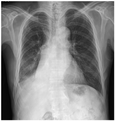
Fig. 1 Chest posterior anterior view shows atelectasis of right middle and lower lobes.
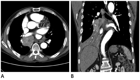
Fig. 2 Chest CT scan. A. Axial image shows 5.8 × 4.8 cm heterogenous enhancing mass obstructing right bronchus intermedius and invading mediastinum. B. Coronal image shows narrowing of right main bronchus by this mass and atelectasis of right middle and lower lobes.
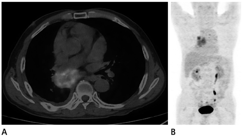
Fig. 3 Positron emission tomography CT scan. A. Axial fusion image shows increased fluorodeoxyglucose (FDG) uptakes (maximal standardized uptake value 6.9) in this mass. B. Torso maximum intensity projection image shows increased FDG uptakes in this mass and mediastinal lymph nodes.
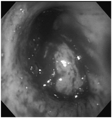
Fig. 4 Bronchoscopy shows protruding round-shape mass in right main bronchus with bloody oozing.
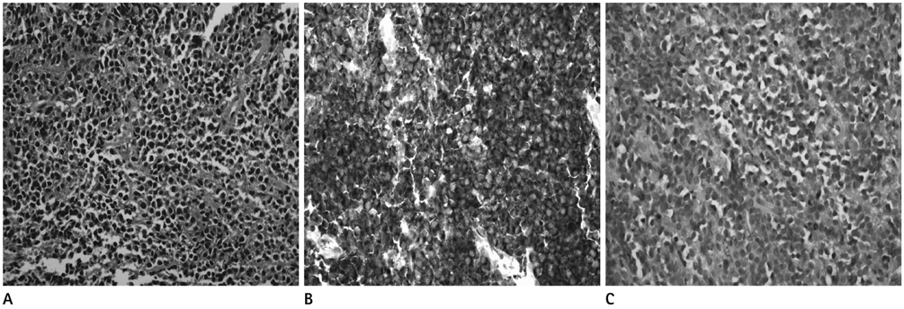
Fig. 5 Microscopic images of tumor tissue sampled by bronchoscopy. A. The tumor composed of sheets of closely packed plasma cells which are characterized by an eccentric nucleus and basophilic cytoplasm. The plasma cells destroy the lung parenchyma and scattered fibrous bands course through the neoplasm (H&E, × 400). B. Immunohistochemically, the tumor cells show diffuse reactivity with monoclonal λ-light chain of the cytoplasm (IHC × 400). C. Immunohistochemically, the tumor cells show negative reaction with κ-light chain (IHC × 400). Note.-IHC = immunohistochemistry
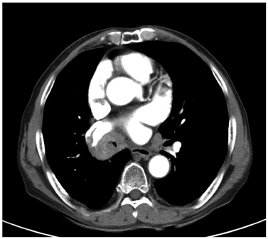
Fig. 6 Follow-up chest CT axial image after 3 months shows decreased size change of this mass and partially opened bronchus intermedius.
Reference
-
1. Montero C, Souto A, Vidal I, Fernández Mdel M, Blanco M, Verea H. [Three cases of primary pulmonary plasmacytoma]. Arch Bronconeumol. 2009; 45:564–566.2. Kim SH, Kim TH, Sohn JW, Yoon HJ, Shin DH, Kim IS, et al. Primary pulmonary plasmacytoma presenting as multiple lung nodules. Korean J Intern Med. 2012; 27:111–113.3. Koss MN, Hochholzer L, Moran CA, Frizzera G. Pulmonary plasmacytomas: a clinicopathologic and immunohisto-chemical study of five cases. Ann Diagn Pathol. 1998; 2:1–11.4. Ujiie H, Okada D, Nakajima Y, Yoshino N, Akiyama H. A case of primary solitary pulmonary plasmacytoma. Ann Thorac Cardiovasc Surg. 2012; 18:239–242.5. Mohammad Taheri Z, Mohammadi F, Karbasi M, Seyfollahi L, Kahkoei S, Ghadiany M, et al. Primary pulmonary plasmacytoma with diffuse alveolar consolidation: a case report. Patholog Res Int. 2010; 2010:463465.6. Lim YH, Park SK, Oh HS, Choi JH, Ahn MJ, Lee YY, et al. A case of primary plasmacytoma of lymph nodes. Korean J Intern Med. 2005; 20:183–186.7. Horiuchi T, Hirokawa M, Oyama Y, Kitabayashi A, Satoh K, Shindoh T, et al. Diffuse pulmonary infiltrates as a roentgenographic manifestation of primary pulmonary plasmacytoma. Am J Med. 1998; 105:72–74.8. Goździuk K, Kedra M, Rybojad P, Sagan D. A rare case of solitary plasmacytoma mimicking a primary lung tumor. Ann Thorac Surg. 2009; 87:e25–e26.
- Full Text Links
-
- Actions
-
Cited
- CITED
-
- Close
- Share
-
- Similar articles
-
- A Case of Extramedullary Plasmacytoma of the Hypopharynx
- A Case of Extramedullary Plasmacytoma of the Tonsil
- A Case of Extramedullary Plasmacytoma of the Larynx
- A Case of Multiple Myeloma with Multiple Intrahepatic Extramedullary Plasmacytomas
- Primary Extramedullary Plasmacytoma of the Colon: A Case Report
