Migration of Sparganosis from the Brain to the Cervical Spinal Cord
- Affiliations
-
- 1Department of Neurosurgery, Hanyang University Guri Hospital, Hanyang University College of Medicine, Guri, Korea. kch5142@hanyang.ac.kr
- KMID: 2018194
- DOI: http://doi.org/10.3340/jkns.2012.51.3.170
Abstract
- Central nervous system (CNS) sparganosis is a rare parasitic infestation caused by ingestion of the raw or inadequately cooked snakes or frogs. Sparganum is well known for its ability of migrating though the tissue, therefore, it can cause various neurological symptoms if it involves neurological systems. A 51-year-old male patient visited our department of neurosurgery complaining of the motor weakness and radiating pain on both upper extremities over 4 months. He had a history of ingesting raw snakes untill his late twenties. The magnetic resonance (MR) images of cervical spine revealed an intramedullary ill-defined enhancing lesion with the aggregated cysts in the upper cervical spinal cord. Under presumptive diagnosis of sparganosis, we took brain MR image. The brain MR images revealed the signal change in right fronto-temporal lobe suggesting the trajectory of parasitic migration via ventricular systems. He underwent a midline myelotomy and granuloma removal followed by the posterior laminoplasty. Pathologic findings showed inflammatory changes and necrosis with keratinized tissue suggesting the CNS sparganosis. We report an uncommon case of CNS sparganosis migrated from the brain to the spinal cord with literature review.
Keyword
MeSH Terms
Figure
-
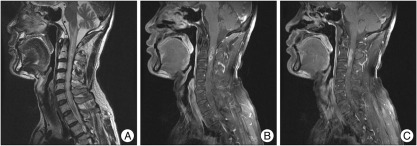
Fig. 1 Cervical magnetic resonance images. A : Irregular high signal intensity is shown from medulla oblongata to C7 level. B and C : Intramedullary ill-defined enhancing lesion with internal multifocal cystic change or aggregated cysts in the upper cervical spinal cord was seen.
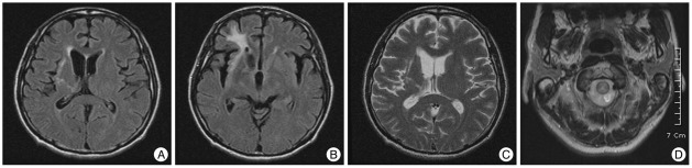
Fig. 2 A : The FLAIR images of MRI show high signal intensity on right fronto-temporal lobe. B and C : A high signal intensity on from basal ganglia and thalamus to ventricle is suggested from the trajectory of parasitic migration via the ventricular system. D : T2-weighted images show high signal intensity on the citerna magna. FLAIR : fluid attenuated inversion recovery.
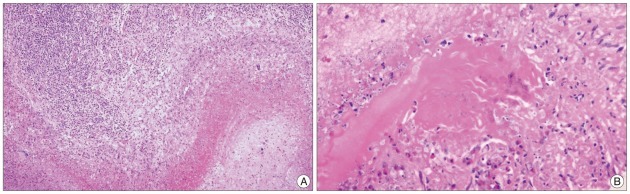
Fig. 3 Photomicrographs are demonstrating the inflammatory change and necrosis with keratinized tissue suggesting granulomatous inflammation (hematoxylin and eosin, A : ×100, B : ×400, respectively).
Reference
-
1. Kim DG, Paek SH, Chang KH, Wang KC, Jung HW, Kim HJ, et al. Cerebral sparganosis : clinical manifestations, treatment, and outcome. J Neurosurg. 1996; 85:1066–1071. PMID: 8929496.2. Kim IY, Jung S, Jung TY, Kang SS, Chung TW. Contralateral migration of cerebral sparganosis through the splenium. Clin Neurol Neurosurg. 2007; 109:720–724. PMID: 17630134.
Article3. Kudesia S, Indira DB, Sarala D, Vani S, Yasha TC, Jayakumar PN, et al. Sparganosis of brain and spinal cord : unusual tapeworm infestation (report of two cases). Clin Neurol Neurosurg. 1998; 100:148–152. PMID: 9746305.
Article4. Ou Q, Li SJ, Cheng XJ. Cerebral sparganosis : a case report. Biosci Trends. 2010; 4:145–147. PMID: 20592465.5. Park JH, Park YS, Kim JS, Roh SW. Sparganosis in the lumbar spine : report of two cases and review of the literature. J Korean Neurosurg Soc. 2011; 49:241–244. PMID: 21607186.
Article6. Rengarajan S, Nanjegowda N, Bhat D, Mahadevan A, Sampath S, Krishna S. Cerebral sparganosis : a diagnostic challenge. Br J Neurosurg. 2008; 22:784–786. PMID: 18661311.
