100 kVp Low-Tube Voltage Abdominal CT in Adults: Radiation Dose Reduction and Image Quality Comparison of 120 kVp Abdominal CT
- Affiliations
-
- 1Department of Radiology, Hallym University Medical Center, Hallym University Sacred Heart Hospital, Anyang, Korea.
- 2Department of Radiology, Soonchunhyang University College of Medicine, Soonchunhyang University Seoul Hospital, Seoul, Korea. hongses@schmc.ac.kr
- KMID: 2353679
- DOI: http://doi.org/10.3348/jksr.2016.75.4.285
Abstract
- PURPOSE
To compare image quality and the effect of radiation dose reduction after decreasing tube voltage from 120 kVp to 100 kVp for abdominal CT in adults.
MATERIALS AND METHODS
A total of 200 patients who underwent abdominal CT at 120 kVp (n = 100) or 100 kVp (n = 100) were enrolled. Automatic tube current modulation was applied with other scan parameters being constant. Radiation dose was calculated based on CT dosimetry index. The image quality of abdominal organs and image noise were assessed quantitatively and qualitatively.
RESULTS
A radiation dose reduction of 13.3% was found in the 100 kVp group. On quantitative analysis, image noise was increased up to 47% in the 100 kVp group. CT numbers of liver, pancreas, renal cortex, aorta, portal vein, and psoas muscle in the 100 kVp group were significantly (p < 0.05) higher than those in the 120 kVp group. Signal-to-noise ratio was significantly higher (p < 0.05) in the 120 kVp group. Contrast-to-noise ratio (CNR) of the liver was higher in the 120 kVp group. However, no significant (p > 0.05) difference was observed in the CNR of other organs between the two groups. On qualitative analysis, noise texture of abdominal organs, artifact, and diagnostic acceptability were not significantly (p > 0.05) different.
CONCLUSION
100 kVp abdominal CT reduced radiation dose by 13.3% without sacrificing image quality compared to 120 kVp abdominal CT.
MeSH Terms
Figure
-

Fig. 1 Body mass index distribution showing no significant difference between 120 kVp and 100 kVp groups (p = 0.9477).

Fig. 2 Quantitative analysis of image noise (mean ± SD). Image noise in the 100 kVp group (10.6 ± 3.8) was higher (p < 0.0001) that that (7.2 ± 1.5) in the 120 kVp group. SD = standard deviation
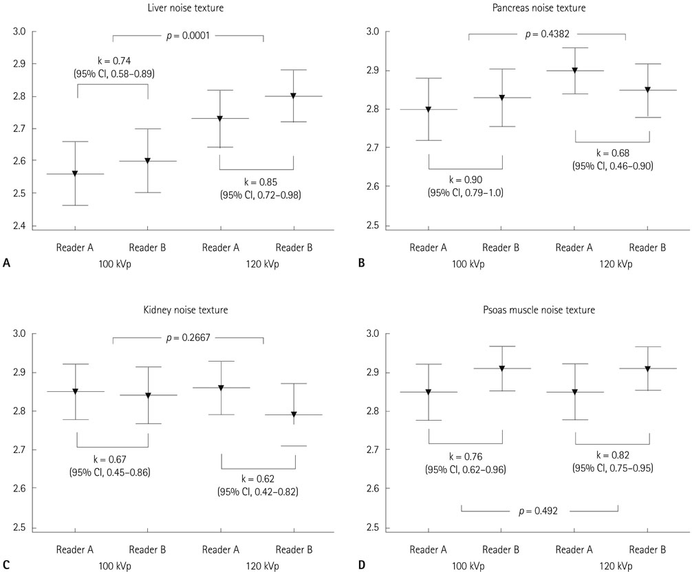
Fig. 3 Qualitative analysis of noise texture (mean ± SD). A. Hepatic noise texture is significantly (p = 0.0001) different between the two groups. B-D. Mean noise texture of pancreas, renal cortex, and psoas muscle is more than 2.8. There is no significant (p > 0.05) difference between the groups. CI = confidence interval, SD = standard deviation, k =interrater agreement
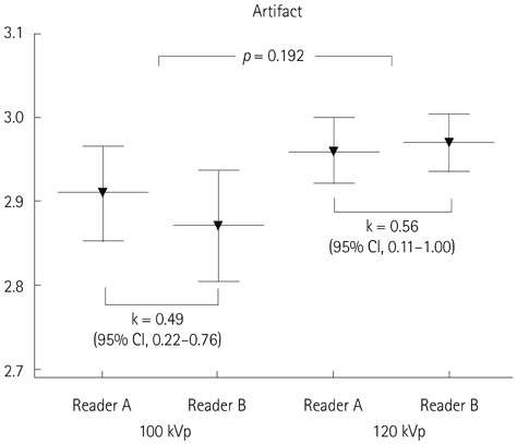
Fig. 4 Qualitative analysis of the artifact (mean ± SD). There is no significant (p = 0.192) difference in artifact rated by two readers between 120 kVp and 100 kVp groups. CI = confidence interval, SD = standard deviation, k = interrater agreement
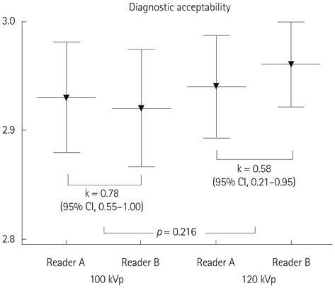
Fig. 5 Qualitative analysis of diagnostic acceptability (mean ± SD). There is no significant (p = 0.216) difference in diagnostic acceptability scored by two readers between 120 kVp and 100 kVp groups. CI = confidence interval, SD = standard deviation, k = interrater agreement
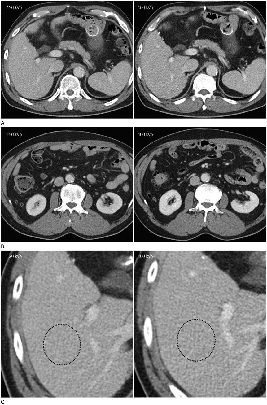
Fig. 6 A 59-year-old male patient underwent gastrectomy of the gastric cancer with post-operative follow-up CT scans using 120 kVp (DLP, 631 mGy·cm) and 100 kVp (DLP, 567 mGy·cm) (window width/level, 45/400) tube voltage at 6-month intervals. A. Hepatic and pancreatic noise texture is rated at 3 points by both readers. SNR and CNR of liver are 19.9 and 8.6 on 120 kVp, respectively, versus 14.5 and 6.8 on 100 kVp. SNR and CNR of pancreas are 17.4 and 6.9 on 120 kVp, respectively, versus 13.2 and 6.0 on 100 kVp. B. SNR and CNR of renal cortex are 30.1 and 19.8 on 120 kVp, respectively, versus 25.9 and 17.8 on 100 kVp. CNR = contrast-to-noise ratio, DLP = dose length product, SNR = signal-to-noise ratio C. On magnification images, hepatic noise texture on 100 kVp shows more pixelated texture (dot-line circle) compared to that of 120 kVp. However, this difference can be hardly detected on normal scale image. The diagnostic acceptability is scored at 3 points by both readers. Estimated DLP reduction is 10.2%. DLP = dose length product
Cited by 1 articles
-
Optimal Phase of Dynamic Computed Tomography for Reliable Size Measurement of Metastatic Neuroendocrine Tumors of the Liver: Comparison between Pre- and Post-Contrast Phases
Jimi Huh, Jisuk Park, Kyung Won Kim, Hyoung Jung Kim, Jong Seok Lee, Jong Hwa Lee, Yoong Ki Jeong, Atul B. Shinagare, Nikhil H. Ramaiya
Korean J Radiol. 2018;19(6):1066-1076. doi: 10.3348/kjr.2018.19.6.1066.
Reference
-
1. Amis ES Jr, Butler PF, Applegate KE, Birnbaum SB, Brateman LF, Hevezi JM, et al. American College of Radiology white paper on radiation dose in medicine. J Am Coll Radiol. 2007; 4:272–284.2. Huda W, Scalzetti EM, Levin G. Technique factors and image quality as functions of patient weight at abdominal CT. Radiology. 2000; 217:430–435.3. McCollough CH, Primak AN, Braun N, Kofler J, Yu L, Christner J. Strategies for reducing radiation dose in CT. Radiol Clin North Am. 2009; 47:27–40.4. Costello JE, Cecava ND, Tucker JE, Bau JL. CT radiation dose: current controversies and dose reduction strategies. AJR Am J Roentgenol. 2013; 201:1283–1290.5. Lee CH, Goo JM, Ye HJ, Ye SJ, Park CM, Chun EJ, et al. Radiation dose modulation techniques in the multidetector CT era: from basics to practice. Radiographics. 2008; 28:1451–1459.6. Schindera ST, Graca P, Patak MA, Abderhalden S, von Allmen G, Vock P, et al. Thoracoabdominal-aortoiliac multidetector-row CT angiography at 80 and 100 kVp: assessment of image quality and radiation dose. Invest Radiol. 2009; 44:650–655.7. Marin D, Nelson RC, Schindera ST, Richard S, Youngblood RS, Yoshizumi TT, et al. Low-tube-voltage, high-tube-current multidetector abdominal CT: improved image quality and decreased radiation dose with adaptive statistical iterative reconstruction algorithm--initial clinical experience. Radiology. 2010; 254:145–153.8. Nakayama Y, Awai K, Funama Y, Hatemura M, Imuta M, Nakaura T, et al. Abdominal CT with low tube voltage: preliminary observations about radiation dose, contrast enhancement, image quality, and noise. Radiology. 2005; 237:945–951.9. Yu L, Li H, Fletcher JG, McCollough CH. Automatic selection of tube potential for radiation dose reduction in CT: a general strategy. Med Phys. 2010; 37:234–243.10. Yu L, Fletcher JG, Grant KL, Carter RE, Hough DM, Barlow JM, et al. Automatic selection of tube potential for radiation dose reduction in vascular and contrast-enhanced abdominopelvic CT. AJR Am J Roentgenol. 2013; 201:W297–W306.11. Eller A, Wuest W, Scharf M, Brand M, Achenbach S, Uder M, et al. Attenuation-based automatic kilovolt (kV)-selection in computed tomography of the chest: effects on radiation exposure and image quality. Eur J Radiol. 2013; 82:2386–2391.12. Heyer CM, Mohr PS, Lemburg SP, Peters SA, Nicolas V. Image quality and radiation exposure at pulmonary CT angiography with 100- or 120-kVp protocol: prospective randomized study. Radiology. 2007; 245:577–583.13. Leschka S, Stolzmann P, Schmid FT, Scheffel H, Stinn B, Marincek B, et al. Low kilovoltage cardiac dual-source CT: attenuation, noise, and radiation dose. Eur Radiol. 2008; 18:1809–1817.14. Waaijer A, Prokop M, Velthuis BK, Bakker CJ, de Kort GA, van Leeuwen MS. Circle of willis at CT angiography: dose reduction and image quality--reducing tube voltage and increasing tube current settings. Radiology. 2007; 242:832–839.15. Siegel MJ, Ramirez-Giraldo JC, Hildebolt C, Bradley D, Schmidt B. Automated low-kilovoltage selection in pediatric computed tomography angiography: phantom study evaluating effects on radiation dose and image quality. Invest Radiol. 2013; 48:584–589.16. Husarik DB, Schindera ST, Morsbach F, Chuck N, Seifert B, Szucs-Farkas Z, et al. Combining automated attenuation-based tube voltage selection and iterative reconstruction: a liver phantom study. Eur Radiol. 2014; 24:657–667.17. Nakaura T, Kidoh M, Nakamura S, Doi Y, Shiraishi S, Awai K, et al. Low-dose abdominal CT protocols with a tube voltage setting of 100 kVp or 80 kVp: performance of radiation dose reduction and influence on visual contrast. Clin Radiol. 2014; 69:804–811.18. Hough DM, Fletcher JG, Grant KL, Fidler JL, Yu L, Geske JR, et al. Lowering kilovoltage to reduce radiation dose in contrast-enhanced abdominal CT: initial assessment of a prototype automated kilovoltage selection tool. AJR Am J Roentgenol. 2012; 199:1070–1077.19. Gnannt R, Winklehner A, Eberli D, Knuth A, Frauenfelder T, Alkadhi H. Automated tube potential selection for standard chest and abdominal CT in follow-up patients with testicular cancer: comparison with fixed tube potential. Eur Radiol. 2012; 22:1937–1945.20. American Association of Physicists in Medicine (AAPM). The measurement, reporting, and management of radiation dose in CT. Report No. 96. College Park, MD: AAPM;2008. July 31, 2014. Available at: http://www.aapm.org/pubs/reports/rpt_96.pdf.21. EUR 16262. European Study Group Website. European guidelines on quality criteria for computed tomography. January 20, 2016. Available at: http://www.drs.dk/guidelines/ct/quality/htmlindex.htm.22. Mayer C, Meyer M, Fink C, Schmidt B, Sedlmair M, Schoenberg SO, et al. Potential for radiation dose savings in abdominal and chest CT using automatic tube voltage selection in combination with automatic tube current modulation. AJR Am J Roentgenol. 2014; 203:292–299.23. Laqmani A, Veldhoen S, Dulz S, Derlin T, Behzadi C, Schmidt-Holtz J, et al. Reduced-dose abdominopelvic CT using hybrid iterative reconstruction in suspected left-sided colonic diverticulitis. Eur Radiol. 2016; 26:216–224.24. Eller A, May MS, Scharf M, Schmid A, Kuefner M, Uder M, et al. Attenuation-based automatic kilovolt selection in abdominal computed tomography: effects on radiation exposure and image quality. Invest Radiol. 2012; 47:559–565.25. Schindera ST, Nelson RC, Yoshizumi T, Toncheva G, Nguyen G, DeLong DM, et al. Effect of automatic tube current modulation on radiation dose and image quality for low tube voltage multidetector row CT angiography: phantom study. Acad Radiol. 2009; 16:997–1002.26. Wintersperger B, Jakobs T, Herzog P, Schaller S, Nikolaou K, Suess C, et al. Aorto-iliac multidetector-row CT angiography with low kV settings: improved vessel enhancement and simultaneous reduction of radiation dose. Eur Radiol. 2005; 15:334–341.27. Schueller-Weidekamm C, Schaefer-Prokop CM, Weber M, Herold CJ, Prokop M. CT angiography of pulmonary arteries to detect pulmonary embolism: improvement of vascular enhancement with low kilovoltage settings. Radiology. 2006; 241:899–907.28. Li X, Yu Y, Liu B, Qian Y, Zhao R. Radiation dose reduction at multidetector CT. Radiology. 2013; 268:925–926.29. Raman SP, Johnson PT, Deshmukh S, Mahesh M, Grant KL, Fishman EK. CT dose reduction applications: available tools on the latest generation of CT scanners. J Am Coll Radiol. 2013; 10:37–41.30. Kalra MK, Woisetschläger M, Dahlström N, Singh S, Lindblom M, Choy G, et al. Radiation dose reduction with sinogram affirmed iterative reconstruction technique for abdominal computed tomography. J Comput Assist Tomogr. 2012; 36:339–346.31. Baker ME, Dong F, Primak A, Obuchowski NA, Einstein D, Gandhi N, et al. Contrast-to-noise ratio and low-contrast object resolution on full- and low-dose MDCT: SAFIRE versus filtered back projection in a low-contrast object phantom and in the liver. AJR Am J Roentgenol. 2012; 199:8–18.32. Ginsburg M, Obara P, Wise L, Wroblewski K, Vannier MW, Dachman AH. BMI-based radiation dose reduction in CT colonography. Acad Radiol. 2013; 20:486–492.
- Full Text Links
-
- Actions
-
Cited
- CITED
-
- Close
- Share
-
- Similar articles
-
- 100 kVp Low-Tube Voltage Abdominal CT in Adults: Radiation Dose Reduction and Image Quality Comparison of 120 kVp Abdominal CT
- Comparison of Image Quality of Shoulder CT Arthrography Conducted Using 120 kVp and 140 kVp Protocols
- A Comparative Study of Image Quality and Radiation Dose with Changes in Tube Voltage and Current for a Digital Chest Radiography
- Windows Setting for Low kVp Abdominal CT: Comparison to 120-kVp CT Images
- 80-kVp CT Using Iterative Reconstruction in Image Space Algorithm for the Detection of Hypervascular Hepatocellular Carcinoma: Phantom and Initial Clinical Experience
