Ductal Carcinoma In Situ of the Breast Arising in Microglandular Adenosis
- Affiliations
-
- 1Department of Radiology, Eulji Hospital, Eulji University School of Medicine, Seoul, Korea. jkan0831@eulji.ac.kr
- 2Department of Surgery, Eulji Hospital, Eulji University School of Medicine, Seoul, Korea.
- 3Department of Pathology, Eulji Hospital, Eulji University School of Medicine, Seoul, Korea.
- KMID: 1439540
- DOI: http://doi.org/10.3348/jksr.2012.67.4.273
Abstract
- Microglandular adenosis of the breast is a benign proliferative lesion, and is a rare subtype of adenosis. The pathologic findings and clinical symptoms can mimic those of breast cancer. Microglandular adenosis has been frequently associated with invasive carcinoma and in situ carcinoma of the breast. Many reports have described a spectrum of the lesions, ranging from microglandular adenosis to cancer arising from microglandular adenosis. However, most of them have focused on the pathology, and there are a few cases that report imaging findings. In the present case, we report the imaging and pathologic findings of a ductal carcinoma in situ arising in microglandular adenosis. A 57-year-old woman detected a palpable mass in her left breast. Mammogram showed an ill-defined irregular isodense mass, and sonogram showed hyperechoic irregular mass with indistinct margin. The patient underwent breast conserving surgery and adjuvant radiotherapy.
MeSH Terms
Figure
-
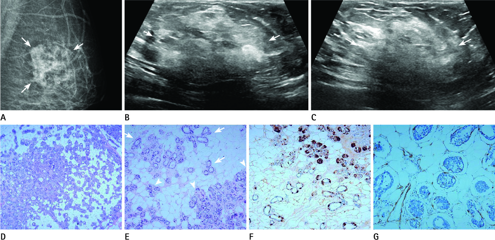
Fig. 1 A 57-year-old woman with a left palpable mass. A. Mammogram shows an ill-defined irregular isodense mass on the left breast (arrows). Some amount of fat is present within the lesion. B, C. Sonogram shows an irregular mass with indistinct margin (arrows). The echogenicity of the mass is predominantly hyperechoic with some heterogeneously isoechoic areas. D. Microscopic finding shows solid type intraductal carcinoma extending to microglandular adenosis (H&E, × 100). E. Transition from uncomplicated microglandular adenosis (arrows) to in situ carcinoma (arrowheads) is clearly noted (H&E, × 200). F. The glands of microglandular adenosis and the tumor cells strongly express S-100 protein (× 200). G. Most of myoepithelial cells are negative for smooth muscle actin on immunohistochemical staining (× 400).
Cited by 1 articles
-
Invasive Breast Carcinoma Arising in Microglandular Adenosis: Two Case Reports
Jung Eun Choi, Young Kyung Bae
J Breast Cancer. 2013;16(4):432-437. doi: 10.4048/jbc.2013.16.4.432.
Reference
-
1. Rosen PP. Adenosis and microglandular adenosis. In : Rosen PP, editor. Rosen's Breast pathology. Philadelphia: Lippincott Williams & Wilkins;2001. p. 152–161.2. Koenig C, Dadmanesh F, Bratthauer GL, Tavassoli FA. Carcinoma arising in microglandular adenosis: an immunohistochemical analysis of 20 intraepithelial and invasive neoplasms. Int J Surg Pathol. 2000; 8:303–315.3. Khalifeh IM, Albarracin C, Diaz LK, Symmans FW, Edgerton ME, Hwang RF, et al. Clinical, histopathologic, and immunohistochemical features of microglandular adenosis and transition into in situ and invasive carcinoma. Am J Surg Pathol. 2008; 32:544–552.4. Rosen PP. Microglandular adenosis. A benign lesion simulating invasive mammary carcinoma. Am J Surg Pathol. 1983; 7:137–144.5. Tavassoli FA, Norris HJ. Microglandular adenosis of the breast. A clinicopathologic study of 11 cases with ultrastructural observations. Am J Surg Pathol. 1983; 7:731–737.6. James BA, Cranor ML, Rosen PP. Carcinoma of the breast arising in microglandular adenosis. Am J Clin Pathol. 1993; 100:507–513.7. Kim DJ, Sun WY, Ryu DH, Park JW, Yun HY, Choi JW, et al. Microglandular adenosis. J Breast Cancer. 2011; 14:72–75.8. Sabaté JM, Gómez A, Torrubia S, Matias-Guiu X, Alonso C, Pericay C, et al. Microglandular adenosis of the breast in a BRCA1 mutation carrier: radiological features. Eur Radiol. 2002; 12:1479–1482.9. Lee YH, Dai YC, Lin IL, Tu CW. Young-aged woman with invasive ductal carcinoma arising in atypical microglandular adenosis: a case report. Pathol Int. 2010; 60:685–689.10. Choi N, Ko ES. Invasive ductal carcinoma in a mammary hamartoma: case report and review of the literature. Korean J Radiol. 2010; 11:687–691.11. Linda A, Zuiani C, Lorenzon M, Furlan A, Girometti R, Londero V, et al. Hyperechoic lesions of the breast: not always benign. AJR Am J Roentgenol. 2011; 196:1219–1224.
- Full Text Links
-
- Actions
-
Cited
- CITED
-
- Close
- Share
-
- Similar articles
-
- Ductal Carcinoma In Situ of the Breast Arising in Microglandular Adenosis
- Mammary Carcinoma Arising in Microglandular Adenosis: A Report of Five Cases
- Invasive Breast Carcinoma Arising in Microglandular Adenosis: Two Case Reports
- Metaplastic Carcinoma with Chondroid Differentiation Arising in Microglandular Adenosis
- Microglandular Adenosis
