A Case Report of Multiple Cavernous Hemangiomas with Fluid-Fluid Levels: A Focus on the Radiologic Features of Dynamic MRI with Subtraction
- Affiliations
-
- 1Department of Radiology, Chung-Ang University Hospital, Seoul, Korea. roentgen@cau.ac.kr
- 2Department of Radiology, Chung-Ang University, Yong-San Hospital, Seoul, Korea.
- KMID: 2097934
- DOI: http://doi.org/10.3348/jksr.2011.64.2.173
Abstract
- Multiple cavernous hemangiomas with fluid-fluid levels were presented in a 40-year-old male patient. These hemangiomas showed nonspecific radiologic features on ultrasound and computed tomography, but gadolinium-enhanced magnetic resonance imaging (MRI) with subtraction revealed fluid-fluid levels with unique and different enhancement of the inferior and superior layer. This is the first case report of cavernous hemangiomas with fluid-fluid levels, containing radiologic features of dynamic MRI with subtraction.
MeSH Terms
Figure
-
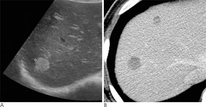
Fig. 1 A 40-year-old man with three hepatic hemangiomas with fluid-fluid levels. A. US shows a well-defined homogeneously hyperechoic nodule in the right lobe of liver. No fluid-fluid levels were demonstrated for all three lesions. B. Dynamic contrast enhanced CT at portal phase shows well-defined low density nodules with layered attenuation differences, which suggest fluid-fluid levels.
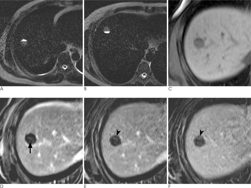
Fig. 2 MRI of the largest hemangioma with a fluid-fluid level. A. Heavily T2-weighted MR image (TR/TE, 1200/462) obtained in the supine position clearly depicts a fluid-fluid level within the hemangioma. B. On prone position, the heavily T2-weighted MR image shows the shift of the fluid-fluid level. C. T1-weighted 3D dynamic MR image without enhancement shows a low signal intensity (SI) nodule with a fluid-fluid level. The superior layer shows lower SI than the inferior layer. D-F. Subtraction MR images obtained 1, 3, and 10 min after injection of contrast medium shows centripetal globular enhancement of the inferior layer (arrow). The superior layer shows subtle enhancement on the 3 min delayed image (arrowhead in E) and gradual filling on the 10 min (arrowhead in F) delayed image. There is a time gap of enhancement between the two layers.
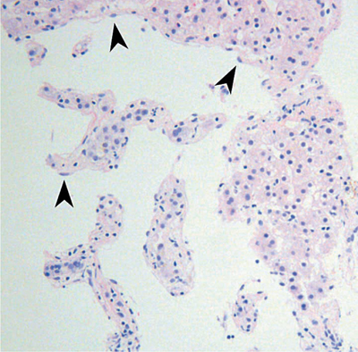
Fig. 3 Photomicrograph shows dilated vascular spaces lined by a single layer of endothelial cells (arrowheads) and separated by connective tissue, which is a typical finding of a cavernous hemangioma (H & E, ×200).
Reference
-
1. Vilgrain V, Boulos L, Vullierme MP, Denys A, Terris B, Menu Y. Imaging of atypical hemangiomas of the liver with pathologic correlation. Radiographics. 2000; 20:379–397.2. Itai Y, Ohtomo K, Kokubo T, Yamauchi T, Okada Y, Makita K. CT demonstration of fluid-fluid levels in nonenhancing hemangiomas of the liver. J Comput Assist Tomogr. 1987; 11:763–765.3. Soyer P, Bluemke DA, Fishman EK, Rymer R. Fluid-fluid levels within focal hepatic lesions: imaging appearance and etiology. Abdom Imaging. 1998; 23:161–165.4. Soyer P, Spelle L, Gouhiri MH, Rondeau Y, Pelage JP, Scherrer A, et al. Gadolinium chelate-enhanced subtraction spoiled gradient-recalled echo MR imaging of hepatic tumors. AJR Am J Roentgenol. 1999; 172:79–82.5. Ward J, Guthrie JA, Scott DJ, Atchley J, Wilson D, Davies MH, et al. Hepatocellular carcinoma in the cirrhotic liver: double-contrast MR imaging for diagnosis. Radiology. 2000; 216:154–162.6. Tung GA, Vaccaro JP, Cronan JJ, Rogg JM. Cavernous hemangioma of the liver: pathologic correlation with high-field MR imaging. AJR Am J Roentgenol. 1994; 162:1113–1117.7. Yamashita Y, Ogata I, Urata J, Takahashi M. Cavernous hemangioma of the liver: pathologic correlation with dynamic CT findings. Radiology. 1997; 203:121–125.8. Obata S, Matsunaga N, Hayashi K, Ohtsubo M, Morikawa T, Takahara O. Fluid-fluid levels in giant cavernous hemangioma of the liver: CT and MRI demonstration. Abdom Imaging. 1998; 23:600–602.9. Ghai S, Dill-Macky M, Wilson S, Haider M. Fluid-fluid levels in cavernous hemangiomas of the liver: baffled? AJR Am J Roentgenol. 2005; 184:3 Suppl. S82–S85.10. Lee J, Lim HK, Jeon YH. Multiple hepatic hemangiomas with fluid-fluid levels. Australas Radiol. 2007; 51:Suppl. B310–B312.
- Full Text Links
-
- Actions
-
Cited
- CITED
-
- Close
- Share
-
- Similar articles
-
- A Case Report of Multiple Cavernous Hemangiomas with Fluid-Fluid Levels: A Focus on the Radiologic Features of Dynamic MRI with Subtraction
- Multiple Cavernous Hemangiomas of the Posterior Mediastinum, Lung, and Liver: A Case Report
- A Case of Spontaneous Intracranial Hypotension: Detection of Cerebrospinal Fluid Leakage by Early Dynamic Radionuclide Cisternography
- Hemodynamics on Three-Phase Dynamic CT and Ultrasonographic Echogenicity of Small Hepatic Cavernous Hemangioma
- Calcified Cavernous Hemangioma of the Ovary: A Case Report
