Progressive Multifocal Leucoencephalopathy Isolated to Posterior Fossa in a Patient with AIDS: DWI and 1H-MRS Features
- Affiliations
-
- 1Department of Radiology, Eulji University Hospital, Daejeon, Korea. midosyu@eulji.ac.kr
- KMID: 2097908
- DOI: http://doi.org/10.3348/jksr.2010.63.5.403
Abstract
- We report a case of progressive multifocal leukoencephalopathy (PML) isolated to the posterior fossa in a 55-year-old male with acquired immune deficiency syndrome(AIDS). Initial MR images revealed a few foci of patchy increased signal intensity(SI)on a T2-weighted image and a diffusion weighted image (DWI) at the pons, right middle cerebellar peduncle, and right cerebellar hemisphere, with no enhancement. After anti-retroviral therapy, follow-up MR images revealed the more prominent extent of previously-seen lesions and newly discovered newly developed focal increased SI on T2-weighted images located left of the inferior cerebellar hemisphere. Proton MR spectroscopy (1H-MRS) showed a slightly increased choline peak (3.2 ppm) and lactate peak (1.35 ppm), as well as a decreased N-acetylaspartate (NAA) peak (2.0 ppm), which suggests active demyelinating disease. DWI and 1H-MRS may support the diagnosis of PML in patients with AIDS.
MeSH Terms
Figure
-

Fig. 1 Initial MRI (A: T2-weighted image (WI), B: T1-WI, C: contrast enhanced T1-WI) revealed a few foci with patchy increased signal intensity (SI) on T2-WI and decreased SI on T1-WI at the pons, right middle cerebellar peduncle, and right cerebellar hemisphere with no enhancement.

Fig. 2 DWI (A) and ADC map (B) showed high signal intensity (SI) on DWI and normal-to-low SI on an ADC map at the peripheral margin of right cerebellar hemisphere, while low SI on DWI and high SI on an ADC map at the center. We suggest that these characteristic features of DWI and the ADC map could be called a "layered phenomenon".

Fig. 3 MR spectroscopy showed a slightly increased choline peak (3.2 ppm) and lactate peak (1.35 ppm), as well as a decreased N-acetylaspartate (NAA) peak (2.0 ppm), suggesting demyelinating disease.
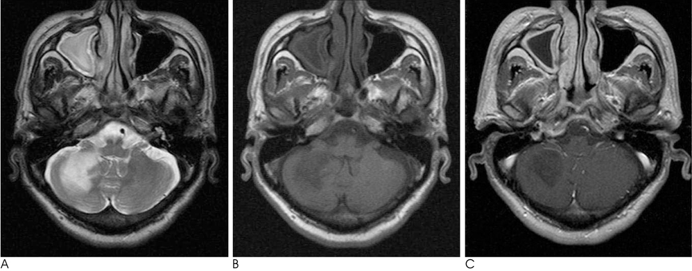
Fig. 4 Twenty-two day follow-up MRI (A: T2-weighted image (WI), B: T1-WI, C: contrast enhanced T1-WI) showed a slight extension of the previous lesions at the pons, right middle cerebellar peduncle, and right cerebellar hemisphere with newly developed (not seen) focal high SI on T2-WI at the left inferior cerebellar hemisphere.
Cited by 2 articles
-
A Case of Progressive Multifocal Leukoencephalopathy in Acquired Immune Deficiency Syndrome Initially Presented with Early Onset Dementia
Pyeong Kang Park, Jung Geun Oh, Seong-Ho Koh, Kyu-Yong Lee, Young Joo Lee, Hojin Choi
Dement Neurocogn Disord. 2014;13(1):20-23. doi: 10.12779/dnd.2014.13.1.20.A Case of Progressive Multifocal Leukoencephalopathy in Acquired Immune Deficiency Syndrome Initially Presented with Early Onset Dementia
Pyeong Kang Park, Jung Geun Oh, Seong-Ho Koh, Kyu-Yong Lee, Young Joo Lee, Hojin Choi
Dement Neurocogn Disord. 2014;13(1):20-23. doi: 10.12779/dnd.2014.13.1.20.
Reference
-
1. Brooks BR, Walker DL. Progressive multifocal leukoencephalopathy. Neurol Clin. 1984; 2:299–313.2. Holman RC, Janssen RS, Buehler JW, Zelasky MT, Hooper WC. Epidermiology of progressive multifocal leukoencephalopathy in the United States: analysis of national mortality and AIDS surveillance data. Neurology. 1991; 41:1733–1736.3. Berger JR, Kaszovitz B, Post MJ, Dickinson G. Progressive multifocal leucoencephalopathy associated with human immunodeficiency virus: a review of the literature with a report of sixteen cases. Ann Intern Med. 1987; 107:78–87.4. Thurnher MM, Post MJ, Rieger A, Kleible-Popov C, Loewe C, Schindker E. Initial and follow-up MR imaging findings in AIDS related progressive multifocal leukoencephalopathy treated with highly active antiretroviral therapy. AJNR Am J Neuroradiol. 2001; 22:977–984.5. Yoon JH, Bang OY, Kim HS. Progressive multifocal leucoencephalopathy in AIDS: proton MR spectroscopy patterns of asynchronous lesions confirmed by serial diffusion-weighted imaging and apparent diffusion coefficient mapping. J Clin Neurol. 2007; 3:200–203.6. Chang L, Ernst T, Tornatore C, Aronow H, Melchor R, Walot I, et al. Metabolite abnormalities in progressive multifocal leukoencephalopathy by proton magnetic resonance spectroscopy. Neurology. 1997; 48:836–845.7. Post MJ, Yiannoutsos C, Simpson D, Booss J, Clifford DB, Cohen B, et al. Progressive multifocal leukoencephalopathy in AIDS: are there any MR findings useful to patient management and predictive of patient survival? AIDS Clinical Trials Group, 243 Team. AJNR Am J Neuroradiol. 1999; 20:1896–1906.8. Whiteman ML, Post MJ, Berger JR, Tate LG, Bell MD, Limonte LP. Progressive multifocal leukoencephalopathy in 47 HIV-seropositive patients: neuroimaging with clinical and pathologic correlation. Radiology. 1993; 187:233–240.9. Kotecha N, George MJ, Smith TW, Corvi F, Litofsky NS. Enhancing progressive multifocal leukoencephalopathy: an indicator of improved immune status. Am J Med. 1998; 105:541–543.
- Full Text Links
-
- Actions
-
Cited
- CITED
-
- Close
- Share
-
- Similar articles
-
- Progressive Multifocal Leucoencephalopathy Isolated to Posterior Fossa in a Patient with AIDS: DWI and 1H-MRS Features
- Progressive Multifocal Leukoencephalopathy in AIDS: Proton MR Spectroscopy Patterns of Asynchronous Lesions Confirmed by Serial Diffusion-Weighted Imaging and Apparent Diffusion Coefficient Mapping
- Serial Follow-up of White Matter Connectivity in a Patient with Progressive Multifocal Leukoencephalopathy Presenting Clinical Improvement
- Changes in Muscular Lipids in Unilateral Isolated Hypertrophy of Gastrocnemius Muscle Can Be Revealed by 1H MR Spectroscopy
- Progressive Multifocal Leukoencephalopathy in the Immunocompromised Patients - 3 Cases Report
