Retroperitoneal Giant Liposarcoma
- Affiliations
-
- 1Department of Urology, Urological Science Institute, Yonsei University Health System, Seoul, Korea. hanwk@yuhs.ac
- 2Department of Pathology, Yonsei University Health System, Seoul, Korea.
- KMID: 1997028
- DOI: http://doi.org/10.4111/kju.2010.51.8.579
Abstract
- Retroperitoneal liposarcoma is an infrequent, locally aggressive malignancy. We report two cases of huge retroperitoneal liposarcomas. The presence of a palpable abdominal mass was a common symptom of the two patients. Preoperative imaging study showed huge retroperitoneal tumors. Both patients underwent complete surgical resections, and a negative microscopic margin was achieved in both cases. The histopathologic diagnosis was a well-differentiated retroperitoneal liposarcoma. Neither of the two patients developed a recurring tumor during the 1.5 years of follow-up.
Keyword
MeSH Terms
Figure
-
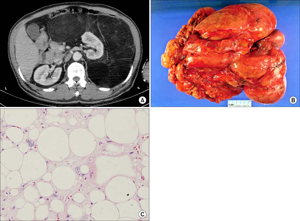
FIG. 1 (A) Computed tomography (CT) findings. The CT image shows a huge, fat-containing mass originating from the retroperitoneum. Encasement of the left kidney by the mass can be observed. (B) Gross findings. The tumor was a multilobulated, huge, mass-like lesion, measuring about 30×30×8 cm. Serial sections of the specimen revealed a homogeneously yellow colored fatty tissue without other significant solid portions. (C) Microscopic findings showing a well-differentiated liposarcoma, composed of mature adipocytes and a few scattered lipoblasts (H&E, ×400).
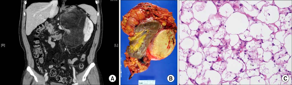
FIG. 2 (A) Computed tomography (CT) image showing a large fatty mass with an enhancing solid portion in the left renal subcapsular area. The kidney shows sprayed. (B) The gross specimen consisting of the left kidney and attached soft tissue, weighing 2,120 g. On bisection, a well-encapsulated, multilobulated, yellowish creamy white solid mass was noted, measuring 18×16×13 cm. Focal necrotic areas are seen, making up 10% of the tumor volume. (C) Microscopic findings showing a well-differentiated liposarcoma and mature-appearing adipose tissue with scattered lipoblasts (H&E, ×200).
Reference
-
1. Perez EA, Gutierrez JC, Moffat FL Jr, Franceschi D, Livingstone AS, Spector SA, et al. Retroperitoneal and truncal sarcomas: prognosis depends upon type not location. Ann Surg Oncol. 2007. 14:1114–1122.2. Seo IY, Won HS, Kim JS, Kim HS, Rim JS. A case of recurrent liposarcoma in retroperitoneum. Korean J Urol. 1994. 35:1375–1378.3. McCallum OJ, Burke JJ 2nd, Childs AJ, Ferro A, Gallup DG. Retroperitoneal liposarcoma weighing over one hundred pounds with review of the literature. Gynecol Oncol. 2006. 103:1152–1154.4. Lewis JJ, Leung D, Woodruff JM, Brennan MF. Retroperitoneal soft-tissue sarcoma: analysis of 500 patients treated and followed at a single institution. Ann Surg. 1998. 228:355–365.5. Choi EH, Yoon JB. Retroperitoneal liposarcoma (pleomorphic type): a case report. Korean J Urol. 1996. 37:1187–1190.6. Bautista N, Su W, O'Connell TX. Retroperitoneal soft-tissue sarcomas: prognosis and treatment of primary and recurrent disease. Am Surg. 2000. 66:832–836.7. Jaques DP, Coit DG, Hajdu SI, Brennan MF. Management of primary and recurrent soft-tissue sarcoma of the retroperitoneum. Ann Surg. 1990. 212:51–59.8. Yol S, Tavli S, Tavli L, Belviranli M, Yosunkaya A. Retroperitoneal and scrotal giant liposarcoma: report of a case. Surg Today. 1998. 28:339–342.9. Shim JE, Lee MY, Kim CM, Jung UY, Na HJ. A case of retroperitoneal liposarcoma. Korean J Urol. 1984. 25:369–371.10. Porter GA, Baxter N, Pisters PW. Retroperitoneal sarcoma: a population-based analysis of epidemiology, surgery, and radiotherapy. Cancer. 2006. 106:1610–1616.
