J Korean Acad Conserv Dent.
2006 Sep;31(5):337-343. 10.5395/JKACD.2006.31.5.337.
Estimation of relation between techniques of dye penetration for microleakage and SEM evaluation for marginal adaptation of the restoration
- Affiliations
-
- 1Department of Conservative Dentistry, College of Dentistry, Dankook University, Cheonan, Korea. donyshin@dankook.ac.kr
- KMID: 1986876
- DOI: http://doi.org/10.5395/JKACD.2006.31.5.337
Abstract
- The purpose of this study was to estimate the relation between techniques used for microleakage from dye penetration and for marginal adaptation from SEM evaluation of the restoration. Using high speed #330 bur, class V cavities (4 x 3 x 1.5 mm around CEJ) were prepared on the buccal surface of 20 extracted human molars. Six dimples as reference points for SEM and dye penetration evaluation were made with 1/2 round bur. Cavity was bulk filled with microhybrid composite resin (Esthet X) and all-in-one adhesive (Xeno III). Teeth were stored in saline solution for one day, after then, they were finished and polished using Sof-Lex system. Fifty percent silver nitrate dye solution was used for the evaluation of microleakage and resin replica was used for marginal adaptation. All of these were done after 1000 times thermocycling between 5 and 55degrees C. Vertical sections were made through three dimples of restoration to obtain samples for the evaluation of dye penetration and inner marginal adaptation. Outer adaptational estimation was done with an intact restoration before sectioning. Dye penetration was determined in three degrees and percentage of outer and inner leaky margin was estimated from SEM image. The data were analysed statistically: Spearman's rho test were used to check relationships between two methods. The result were as follows: 1. There were significant relationships between degree of dye penetration and inner and outer marginal adaptations each (p < 0.01). 2. However, there was no significant relationship between the results of inner and outer marginal adaptation. Within the results of this study, relationship between the percentage of marginal adaptation and microleakage shows significant relationship. However, inner and outer marginal adaptation did not show any significant relationship mutually.
Keyword
MeSH Terms
Figure
-
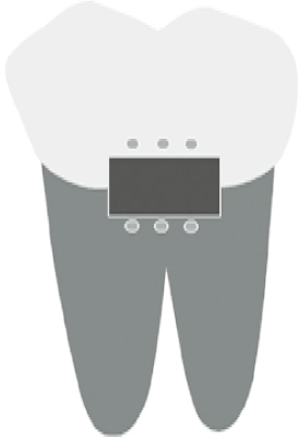
Figure 1 Schematic drawing of cavity and hemi-sphere indents.
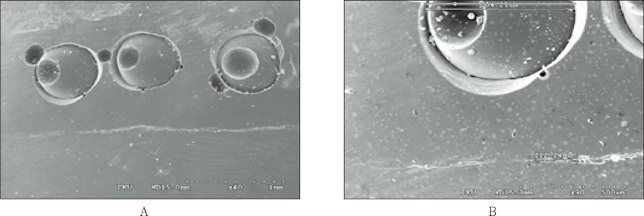
Figure 2 Outer marginal adaptation from SEM evaluation: SEM image of outer marginal adaptation (A), mesuring length of full outer margin and marginal gap (B).
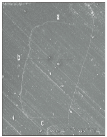
Figure 3 Inner marginal adaptation from SEM evaluation (× 40): a-length of occlusal wall: b-length of axial wall: c-length of gingival wall.
Cited by 1 articles
-
Comparison of marginal microleakage between low and high flowable resins in class V cavity
Sang-Bae Bae, Young-Gon Cho, Myeong-Seon Lee
J Korean Acad Conserv Dent. 2009;34(6):477-483. doi: 10.5395/JKACD.2009.34.6.477.
Reference
-
1. Saboia Vde P, Pimenta LA, Ambrosano GM. Effect of collagen removal on microleakage of resin composite restoration. Oper Dent. 2002. 27(1):38–43.2. Adamo HL, Buruiana R, Schertzer L, Boylan RJ. A comparison of MTA, Super-EBA, composite and amalgam as root filling materials using a bacterial microleakage model. Int Endod J. 1999. 32:197–203.
Article3. Hembree JH Jr, Andrews JT. Microleakage of several Class V anterior restorative materials: a laboratory study. J Am Dent Assoc. 1978. 97(2):179–183.
Article4. Manhart J, Chen HY, Mehl A, Weber K, Hickel R. Marginal quality and microleakage of adhesive class V restorations. J Dent. 2001. 29(2):123–130.
Article5. Youngson CC, Jones JC, Fox K, Smith IS, Wood DJ, Gale M. A fluid filteration and clearing technique to assess microleakage associated with three dentine bonding systems. J Dent. 1999. 27(3):223–233.
Article6. Von Fraunhofer JA, Adachi EI, Barnes DM, Romberg E. The effect of tooth preparation on microleakage behavior. Oper Dent. 2000. 25(6):526–533.7. Gale MS, Darvell BW, Cheung GS. Three-dimensional reconstruction of microleakage pattern using a sequential grinding technique. J Dent. 1994. 22(6):370–375.
Article8. Ha SY, Shin DH. New quantitative measuring technique for microleakage of the restored tooth through 3D reconstruction. J Korean Acad Conserv Dent. 2004. 29(5):413–422.
Article9. Cho KM, Shin DH. Quantitiative evaluation of microleakage using microtomography in class V restorations. 2004. Dankook University;MS thesis.10. Santini A, Mitchell S. Microleakage of composite restorations bonded with three new dentin bonding agents. J Esthet Dent. 1998. 10(6):296–304.
Article11. Youngson CC, Glyn Jones JC, Manogue M, Smith IS. In vitro dentinal penetration by tracers used in microleakage studies. Int Endod J. 1998. 31(2):90–99.12. Gladys S, Van Meerbeek B, Lambrechts P, Vanherle G. Microlekeage of adhesive restorative materials. Am J Dent. 2001. 14(3):170–176.13. Pashley DH, Depew DD. Effects of the smear layer, copalite and oxalate on microleakage. Oper Dent. 1986. 11:95–102.14. Crim GA, Chapman KW. reducing microleakage in class II restorations: an in vitro study. Quintessence Int. 1994. 25(11):781–785.15. Dietschi D, Bindi G, Krejci I, Davidson C. Marginal and internal adaptation of stratified compomer-composite class II restorations. Oper Dent. 2002. 27:500–509.16. Alhadainy HA, Elsaed HY, Elbaghdady YM. An electrochemical study of the sealing ability of different retrofilling materials. J Endod. 1993. 19(10):508–511.
Article17. Iwami Y, Yamamoto H, Ebisu S. A new electrical method for detecting marginal leakage of in vitro resin restorations. J Dent. 2000. 28(4):241–247.
Article18. Manhart J, Chen HY, Mehl A, Weber K, Hickel R. marginal quality and microleakage of adhesive class V restorations. J Dent. 2001. 29(2):123–130.
Article19. Roulet JF. Marginal integrity, clinical significance. J Dent. 1994. 22:suppl 1. S9–S12. Review.
Article20. Gladys S, Van Meerbeek B, Inokoshi S, Willems G, Braem M, Lambrechts P, Vanherle G. Clinical and semiquantitative marginal analysis of four toothcoloured inlay systems at 3 years. J Dent. 1995. 23(6):329–338.
Article21. Miyazaki M, Onose H, Moore BK. Effect of operator variability on dentin bond strength of two-step bonding systems. Am J Dent. 2000. 13(2):101–104.22. Yoshiyama M, Carvalho R, Sano H, Honer J, Brewer PD, Pashley DH. Regional bond strengths of resins to human root dentine. J Dent. 1996. 24:435–442.
Article23. Kazemi RB, Meiers JC, Peppers K. Effect of caries disclosing agents on bond strengths of total-etch and self-etching primer dentin bonding systems to resin composite. Oper Dent. 2002. 27:238–242.24. Feilzer AJ, DeGee AJ, Davidson CL. Curing contraction of composites and glass-ionomer cements. J Prosthet Dent. 1988. 59:297–300.
Article25. Feilzer AJ, deGeeAJ , Davidson CL. Setting stress in composite resin in relation to the configuration of the restoration. J Dent Res. 1987. 66:1636–1639.
Article26. Santini A, Plasschehaert AJM, Mitchell S. Effect of composite resin placement techniques on the microleakage of two self-etching dentin-bonding agents. Am J Dent. 2001. 14(3):132–136.
- Full Text Links
-
- Actions
-
Cited
- CITED
-
- Close
- Share
-
- Similar articles
-
- New quantitative measuring technique for microleakage of the restored tooth through 3D reconstruction
- Microleakage of the class V cavity according to restoration site and cavity size using SEM and three-dimensional reconstruction techniques
- Microhardness and microleakage of composite resin cured by visible light with various band of wavelength
- Mechanical properties and microleakage of composite resin materials cured by variable light intensities
- Marginal microleakage of cervical composite resin restorations bonded using etch-and-rinse and self-etch adhesives: two dimensional vs. three dimensional methods
