Anatomical variations of trabecular bone structure in intraoral radiographs using fractal and particles count analyses
- Affiliations
-
- 1Department of Oral Radiology, Faculty of Dentistry, Minia University, El Menia, Egypt.
- 2Department of Oral and Maxillofacial Radiology and Dental Research Institute, School of Dentistry, Seoul National University, Seoul, Korea. hmslsh@snu.ac.kr
- 3Division of Oral and Maxillofacial Radiology, Department of Periodontics and Oral Medicine, School of Dentistry, University of Michigan, Ann Arbor, USA.
- KMID: 1974401
- DOI: http://doi.org/10.5624/isd.2012.42.1.5
Abstract
- PURPOSE
This study was performed to evaluate possible variations in maxillary and mandibular bone texture of normal population using the fractal analysis, particles count, and area fraction in intraoral radiographs.
MATERIALS AND METHODS
Periapical radiographs of patients who had full mouth intraoral radiographs were collected. Regions of interest (100x100 pixels) were located between the teeth of the maxillary anterior, premolar, and molar area, as well as the mandibular anterior, premolar, and molar areas. The fractal dimension (FD) was calculated by using the box counting method. The particle count (PC) and area fraction (AF) analyses were also performed.
RESULTS
There was no significant difference in the FD values among the different groups of age, gender, upper, and lower jaws. The mean FD value was 1.49+/-0.01. The mean PC ranged from 44 to 54, and the mean AF ranged from 10.92 to 11.85. The values of FD, PC, and AF were significantly correlated with each other except for the upper molar area.
CONCLUSION
According to the results, patients with normal trabecular pattern showed a FD of approximately 1.5. Based on these results, further investigation would be recommended if the FD value of patient significantly differenct from this number, since the alteration of this value indicates microstructural modification of trabecular pattern of the jaws. Additionally, with periapical radiographs, simple and cost-effective, PC and AF could be used to assess the deviation from the normal.
Figure
-
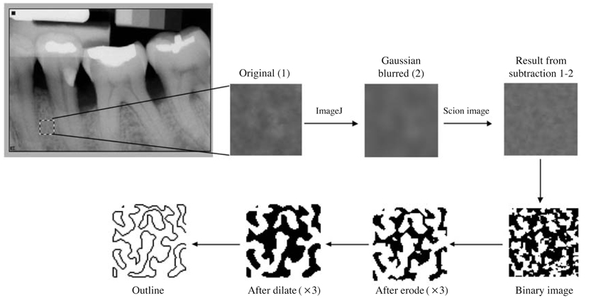
Fig. 1 Image processing procedure to make the outline image from a periapical radiograph. The resultant image is used for the analyses of fractal dimension, particles count, and area fraction.
Cited by 4 articles
-
Method for Automated Selection of the Trabecular Area in Digital Periapical Radiographic Images Using Morphological Operations
Enny Itje Sela, Reza Pulungan, Rini Widyaningrum, Rurie Ratna Shantiningsih
Healthc Inform Res. 2019;25(3):193-200. doi: 10.4258/hir.2019.25.3.193.The three-dimensional microstructure of trabecular bone: Analysis of site-specific variation in the human jaw bone
Jo-Eun Kim, Jae-Myung Shin, Sung-Ook Oh, Won-Jin Yi, Min-Suk Heo, Sam-Sun Lee, Soon-Chul Choi, Kyung-Hoe Huh
Imaging Sci Dent. 2013;43(4):227-233. doi: 10.5624/isd.2013.43.4.227.Prediction of age-related osteoporosis using fractal analysis on panoramic radiographs
Kwang-Joon Koh, Ha-Na Park, Kyoung-A Kim
Imaging Sci Dent. 2012;42(4):231-235. doi: 10.5624/isd.2012.42.4.231.Impact of radiotherapy on mandibular bone: A retrospective study of digital panoramic radiographs
Luiz Felipe Palma, Ricardo Yudi Tateno, Cíntia Maria Remondes, Marcelo Marcucci, Arthur Rodriguez Gonzalez Cortes
Imaging Sci Dent. 2020;50(1):31-36. doi: 10.5624/isd.2020.50.1.31.
Reference
-
1. Jett S, Shrout MK, Mailhot JM, Potter BJ, Borke JL. An evaluation of the origin of trabecular bone patterns using visual and digital image analysis. Oral Surg Oral Med Oral Pathol Oral Radiol Endod. 2004. 98:598–604.2. Lang P, Steiger P, Faulkner K, Gluer C, Genant HK. Osteoporosis. Current techniques and recent developments in quantitative bone densitometry. Radiol Clin North Am. 1991. 29:49–76.3. Bender IB, Seltzer S. Roentgenographic and direct observation of experimental lesions in bone: I. 1961. J Endod. 2003. 29:702–706.
Article4. Parfitt AM. Trabecular bone architecture in the pathogenesis and prevention of fracture. Am J Med. 1987. 82:68–72.
Article5. Genant HK, Cooper C, Poor G, Reid I, Ehrlich G, Kanis J, et al. Interim report and recommendations of the World Health Organization Task-Force for Osteoporosis. Osteoporos Int. 1999. 10:259–264.
Article6. Otis LL, Hong JS, Tuncay OC. Bone structure effect on root resorption. Orthod Craniofac Res. 2004. 7:165–177.
Article7. Heo MS, Park KS, Lee SS, Choi SC, Koak JY, Heo SJ, et al. Fractal analysis of mandibular bony healing after orthognathic surgery. Oral Surg Oral Med Oral Pathol Oral Radiol Endod. 2002. 94:763–767.8. Yaşar F, Akgünlü F. The differences in panoramic mandibular indices and fractal dimension between patients with and without spinal osteoporosis. Dentomaxillofac Radiol. 2006. 35:1–9.9. Prouteau S, Ducher G, Nanyan P, Lemineur G, Benhamou L, Courteix D. Fractal analysis of bone texture: a screening tool for stress fracture risk? Eur J Clin Invest. 2004. 34:137–142.10. Chen SK, Oviir T, Lin CH, Leu LJ, Cho BH, Hollender L. Digital imaging analysis with mathematical morphology and fractal dimension for evaluation of periapical lesions following endodontic treatment. Oral Surg Oral Med Oral Pathol Oral Radiol Endod. 2005. 100:467–472.11. Shrout MK, Jett S, Mailhot JM, Potter BJ, Borke JL, Hildebolt CF. Digital image analysis of cadaver mandibular trabecular bone patterns. J Periodontol. 2003. 74:1342–1347.
Article12. Parkinson IH, Fazzalari NL. Methodological principles for fractal analysis of trabecular bone. J Microsc. 2000. 198:134–142.
Article13. Ergun S, Saracoglu A, Guneri P, Ozpinar B. Application of fractal analysis in hyperparathyroidism. Dentomaxillofac Radiol. 2009. 38:281–288.
Article14. White SC, Rudolph DJ. Alterations of the trabecular pattern of the jaws in patients with osteoporosis. Oral Surg Oral Med Oral Pathol Oral Radiol Endod. 1999. 88:628–635.
Article15. White SC, Rudolph DJ, Ma L. Influence of x-ray beam angulation and exposure on morphologic features of trabecular bone. Int J Oral Biol. 1999. 24:17–23.16. Yasar F, Akgünlü F. Fractal dimension and lacunarity analysis of dental radiographs. Dentomaxillofac Radiol. 2005. 34:261–267.17. Haire TJ, Hodgskinson R, Ganney PS, Langton CM. A comparison of porosity, fabric and fractal dimension as predictors of the Young's modulus of equine cancellous bone. Med Eng Phys. 1998. 20:588–593.
Article18. Buckland-Wright JC, Lynch JA, Rymer J, Fogelman I. Fractal signature analysis of macroradiographs measures trabecular organization in lumbar vertebrae of postmenopausal women. Calcif Tissue Int. 1994. 54:106–112.
Article19. Lynch JA, Hawkes DJ, Buckland-Wright JC. Analysis of texture in macroradiographs of osteoarthritic knees using fractal signature. Phys Med Biol. 1991. 36:709–722.20. Lynch JA, Hawkes DJ, Buckland-Wright JC. A robust and accurate method for calculating fractal signature of texture in macroradiographs of osteoarthritic knees. Med Inform (Lond). 1991. 16:241–251.21. Fazzalari NL, Parkinson IH. Fractal properties of cancellous bone of the iliac crest in vertebral crush fracture. Bone. 1998. 23:53–57.
Article22. Haidekker MA, Andresen R, Evertsz CJ, Banzer D, Peitgen HO. Assessing the degree of osteoporosis in the axial skeleton using the dependence of the fractal dimension on the grey level threshold. Br J Radiol. 1997. 70:586–593.
Article23. Dey P, Rajesh L. Fractal dimension in endometrial carcinoma. Anal Quant Cytol Histol. 2004. 26:113–116.
Article24. Jolley L, Majumdar S, Kapila S. Technical factors in fractal analysis of periapical radiographs. Dentomaxillofac Radiol. 2006. 35:393–397.
Article25. Demirbas AK, Ergün S, Güneri P, Aktener OP, Boyacioğglu H. Mandibular bone changes in sickle cell anemia: fractal analysis. Oral Surg Oral Med Oral Pathol Oral Radiol Endod. 2008. 106:e41–e48.
Article26. Shrout MK, Roberson B, Potter BJ, Mailhot JM, Hildebolt CF. A comparison of 2 patient populations using fractal analysis. J Periodontol. 1998. 69:9–13.
Article27. Chappard C, Brunet-Imbault B, Lemineur G, Giraudeau B, Basillais A, Harba R, et al. Anisotropy changes in post-menopausal osteoporosis: characterization by a new index applied to trabecular bone radiographic images. Osteoporos Int. 2005. 16:1193–1202.28. Southard TE, Southard KA, Lee A. Alveolar process fractal dimension and postcranial bone density. Oral Surg Oral Med Oral Pathol Oral Radiol Endod. 2001. 91:486–491.29. Ruttimann UE, Webber RL, Hazelrig JB. Fractal dimension from radiographs of peridental alveolar bone. A possible diagnostic indicator of osteoporosis. Oral Surg Oral Med Oral Pathol. 1992. 74:98–110.
Article30. Southard TE, Southard KA, Jakobsen JR, Hillis SL, Najim CA. Fractal dimension in radiographic analysis of alveolar process bone. Oral Surg Oral Med Oral Pathol Oral Radiol Endod. 1996. 82:569–576.
Article
- Full Text Links
-
- Actions
-
Cited
- CITED
-
- Close
- Share
-
- Similar articles
-
- The influence of X ray beam angulation on the fractal analysis of trabecular architecture in human dry mandible using standardized tile counting method
- Characterization of trabecular bone structure using 2D Fourier transformation and fractal analysis
- Changes in the fractal dimension of peri-implant trabecular bone after loading: a retrospective study
- Structural complexity of the craniofacial trabecular bone in multiple myeloma assessed by fractal analysis
- Prediction of osteoporosis using fractal analysis on periapical and panoramic radiographs
