Percutaneous Endoscopic Discectomy for Lumbar Disc Herniation
- Affiliations
-
- 1Department of Orthopedic Surgery, Chonnam National University Hospital, Gwangju, Korea. sanggunlee@lycos.co.kr
- KMID: 1941656
- DOI: http://doi.org/10.4184/jkss.2007.14.3.212
Abstract
- Percutaneous endoscopic lumbar discectomy is a widely used procedure. In addition to the surgical techniques, the proper selection of the patients and appropriate approaching portal is important improving the clinical results. The choice of the approaching portal is related to the distance of migration and spinal canal encroachment in addition to the type of herniation type. In addition, it is essential to know the anatomic characteristics at each level of the lumbar spine in addition to the indications of the various approaching portals.
MeSH Terms
Figure
-
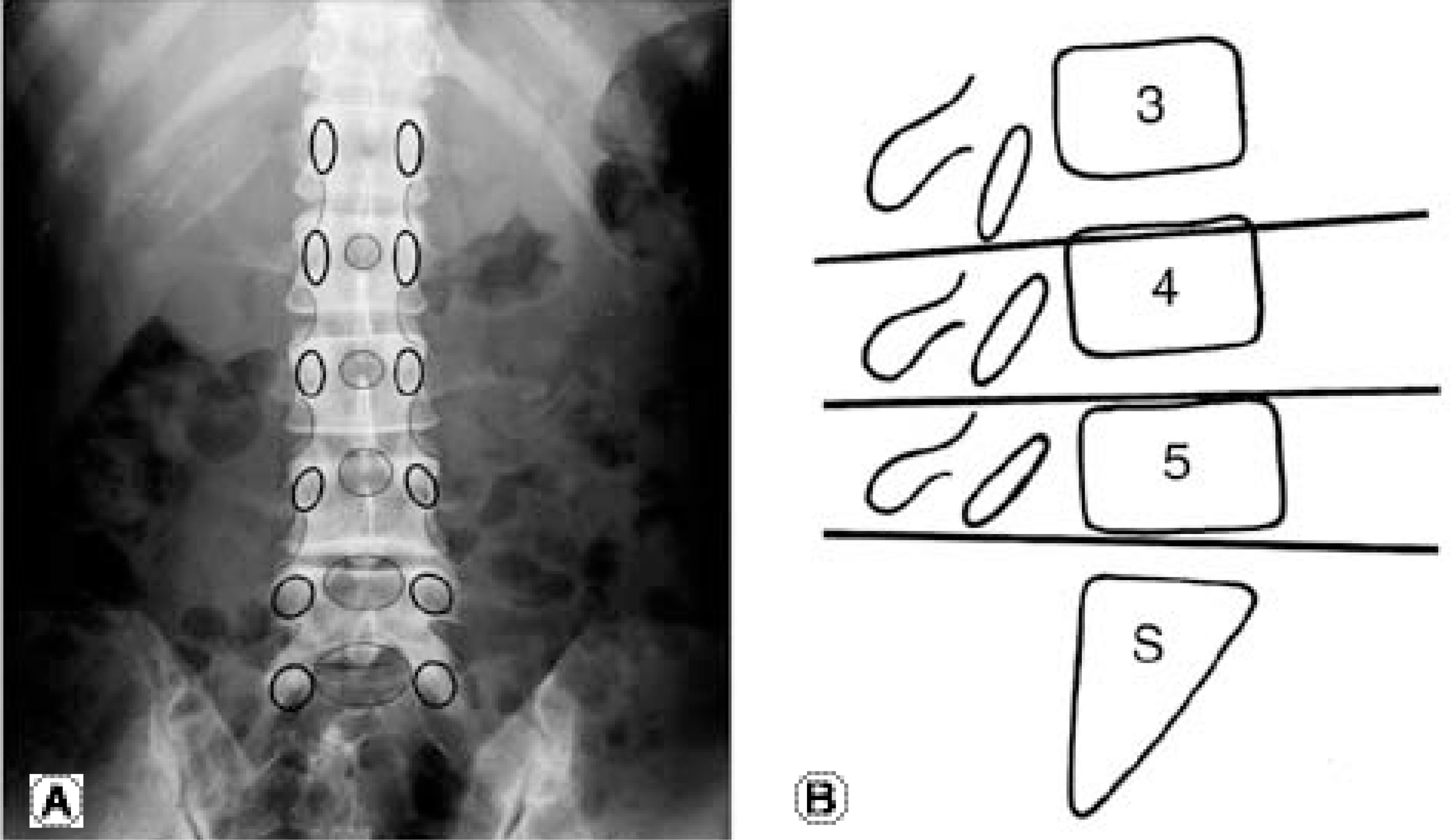
Fig. 1. Anatomical characteristics of lumbar spine (A) Relationships of the interpedicular distance, interlaminar space, and laminar overlap at each lumbar segment. (B) Schematic description of laminar overhanging
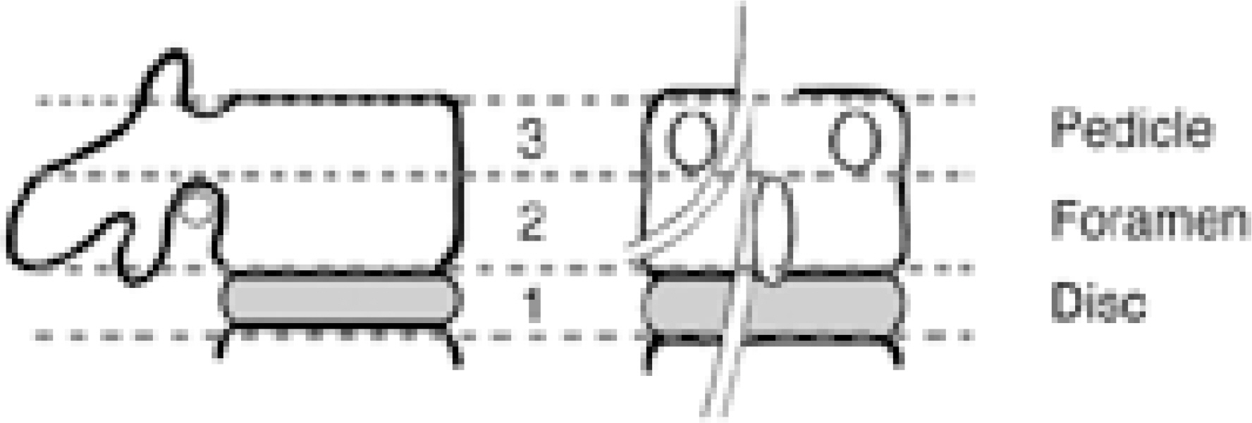
Fig. 2. Schematic diagram of the ‘Three-storied anatomical house concept’.
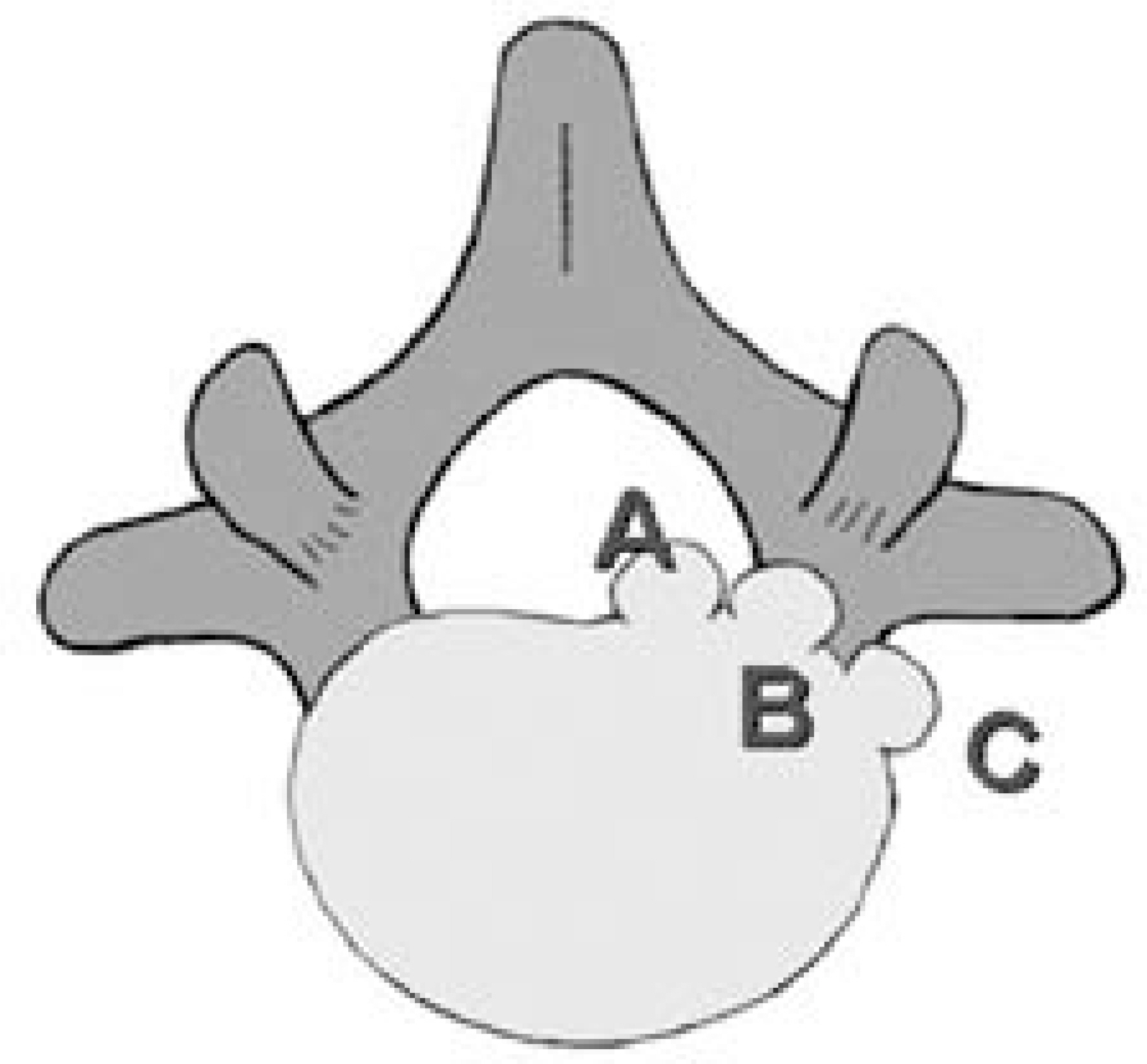
Fig. 3. Classification of the herniated disc related to the axial plane. A-intraspinal, B-foraminal, C-extraforaminal
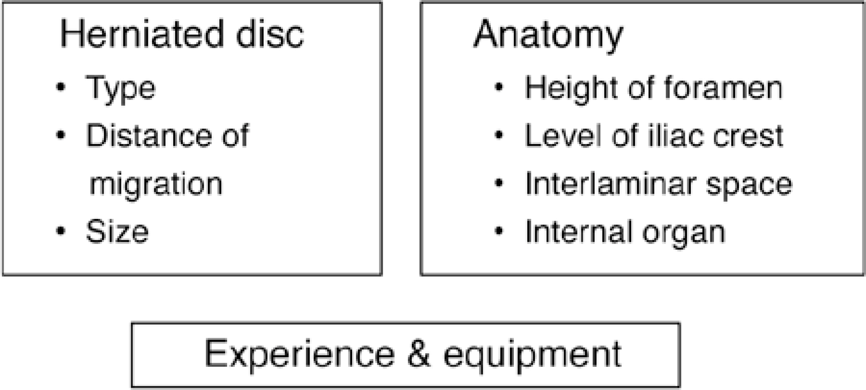
Fig. 4. Considerations to select endoscopic approaching portals.
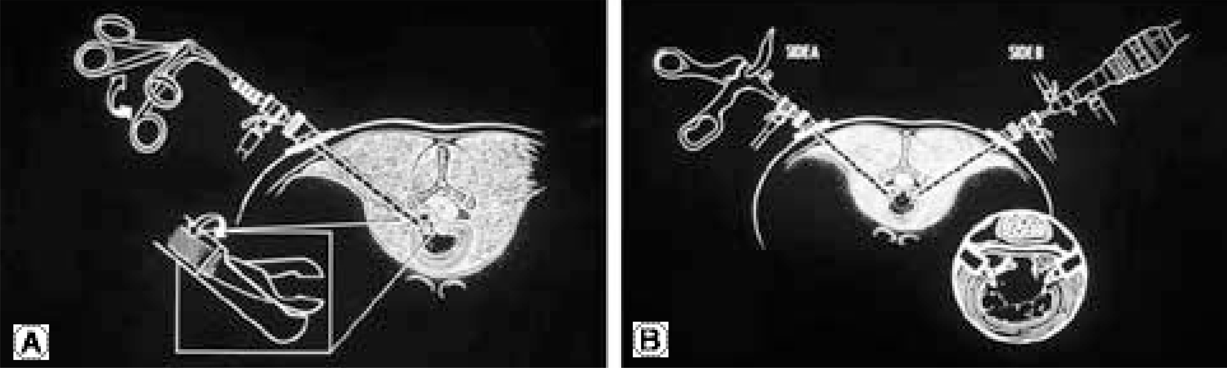
Fig. 5. Posterolateral approaching portal (A) uniportal method (B) biportal method
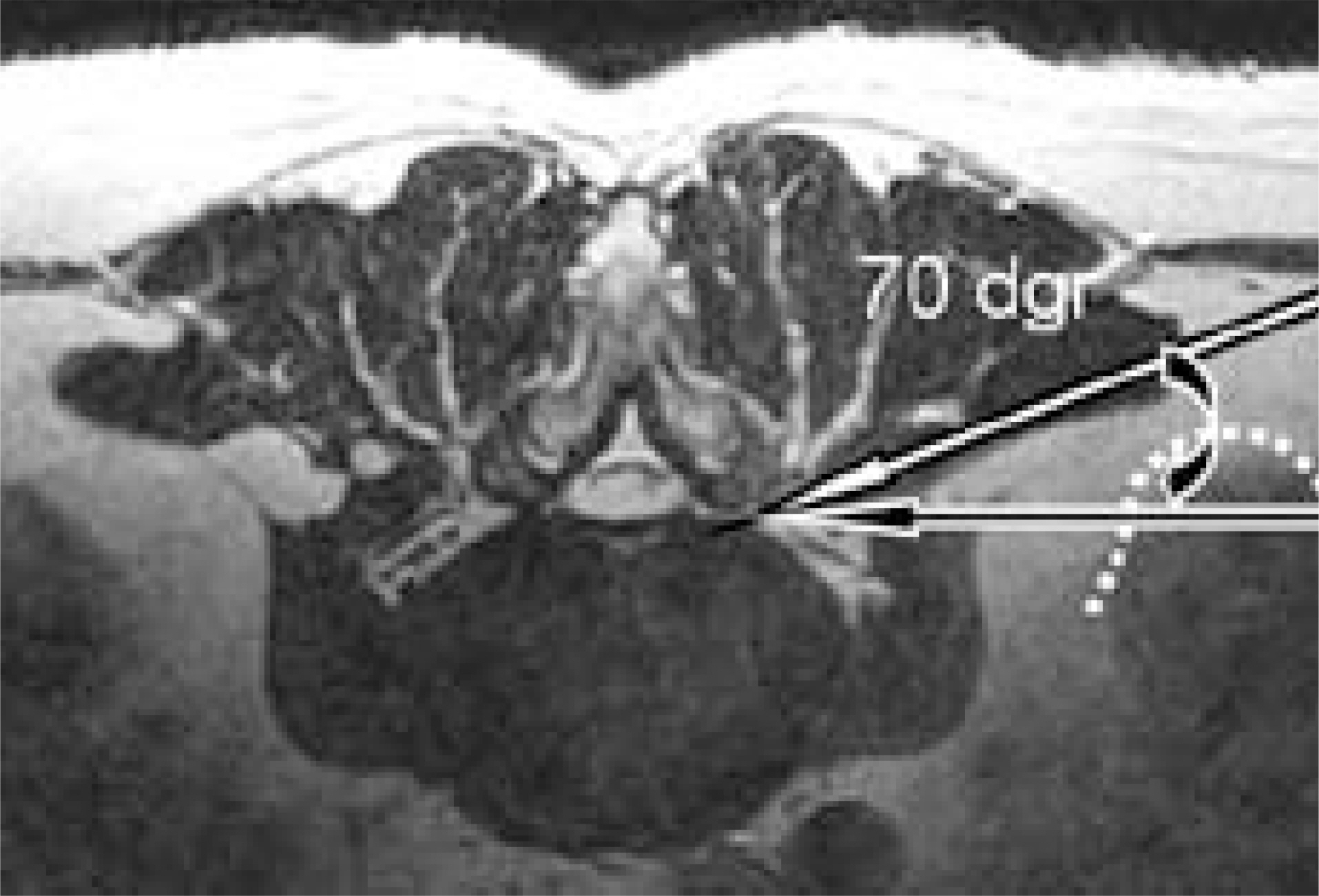
Fig. 6. Insertion angle of the transforaminal approaching portal not to injure the internal organ.
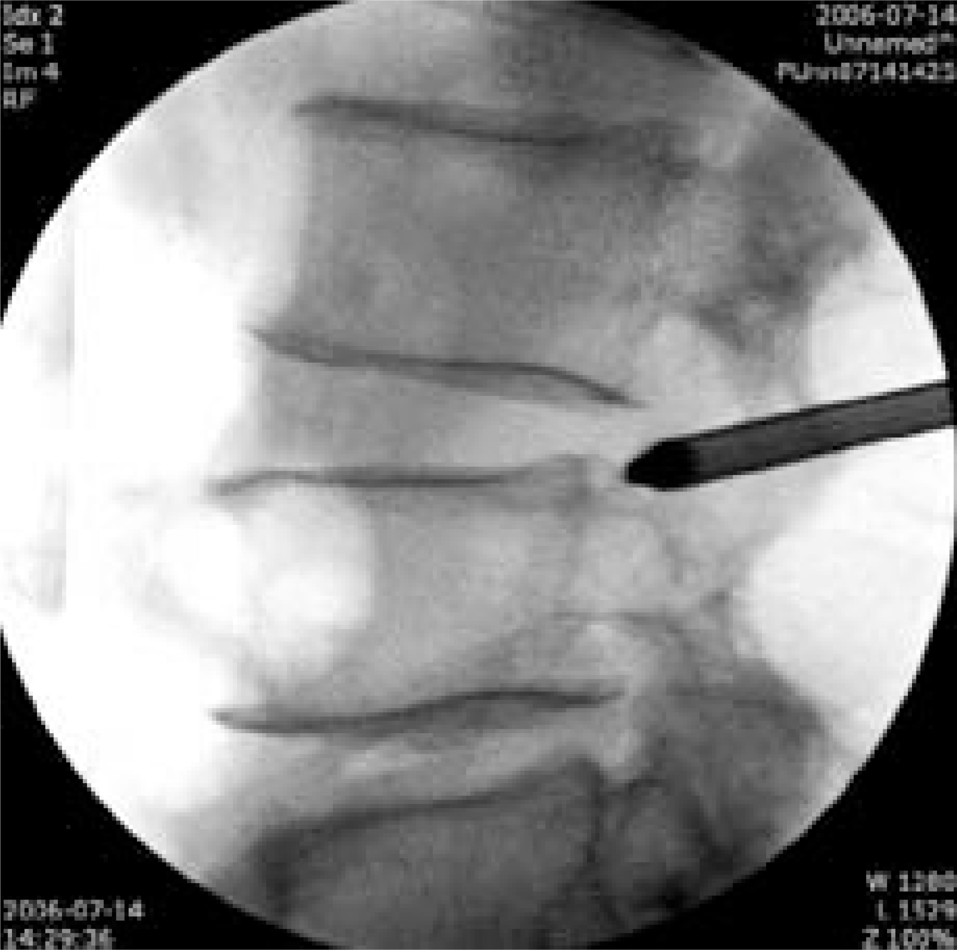
Fig. 7. Fluoroscopic image shows the obturator located just above superior wall of pedicle to avoid injury of the exit root.
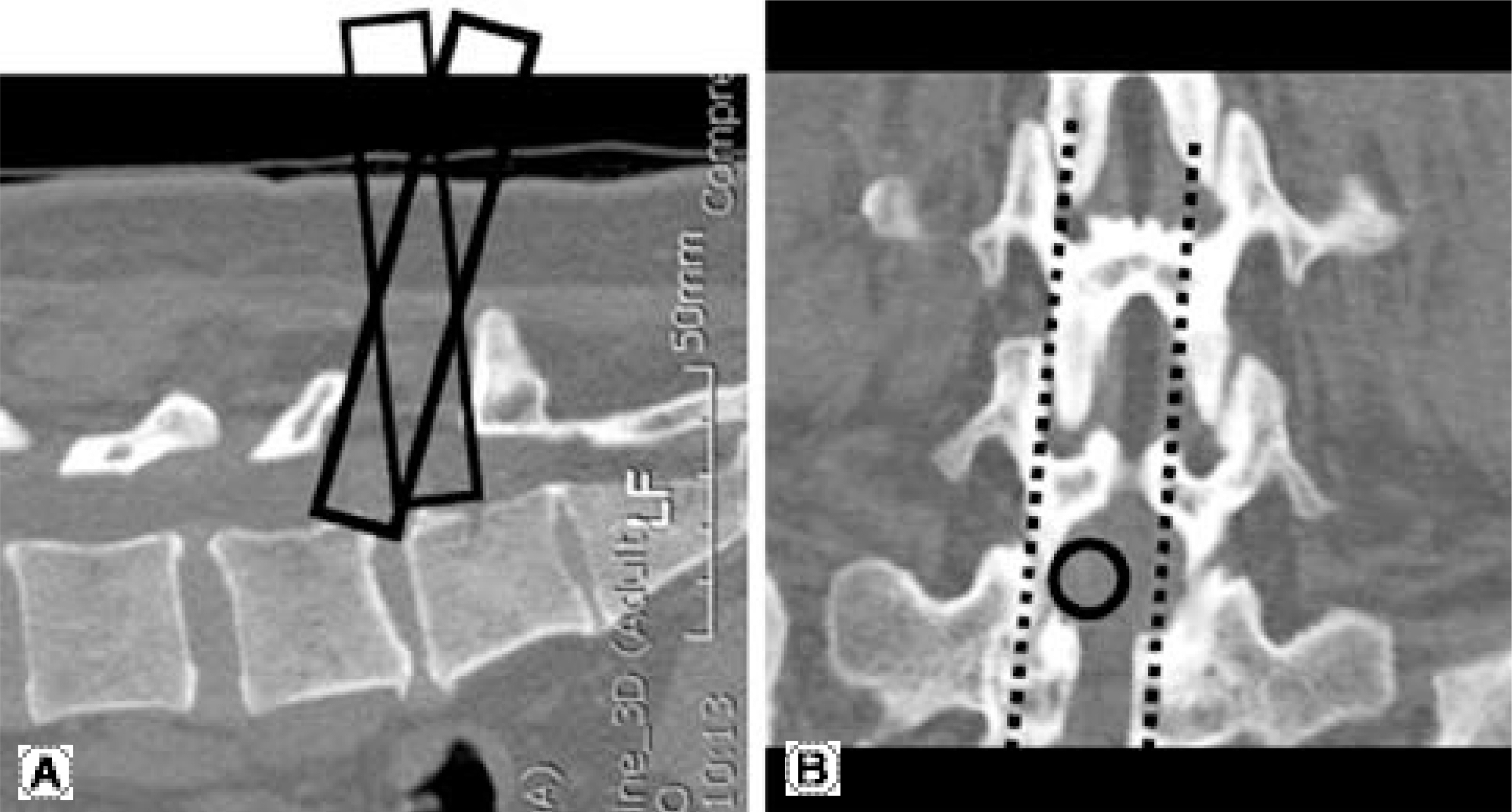
Fig. 8. Sagittal (A) and coronal (B) reformatting images of the CT scan show the enough interlaminar space at L5-S1.
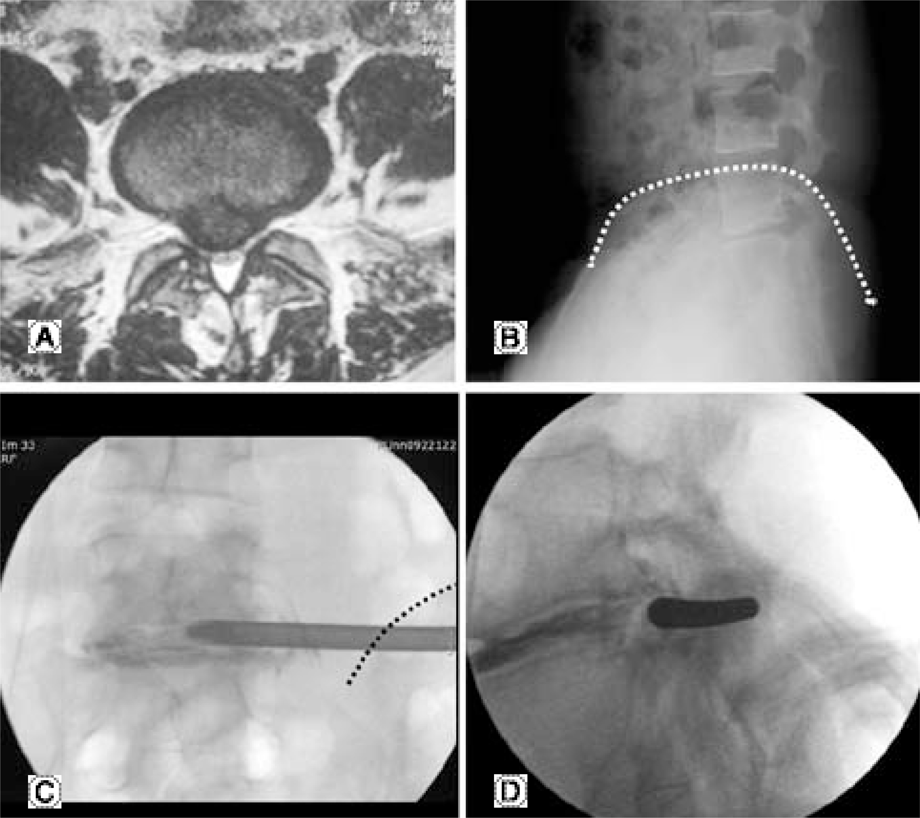
Fig. 9. Transiliac transforaminal approaching portal. (A) Axial T2-weighted MR image shows huge central disc herniaton related with bilateral symptoms. (B) Relationship between the iliac crest and neural foramen of L5-S1. (C, D) Intraoperative fluoroscopic images after insertion of cannula through the transiliac osseous tunnel. The dotted line represented an iliac crest.
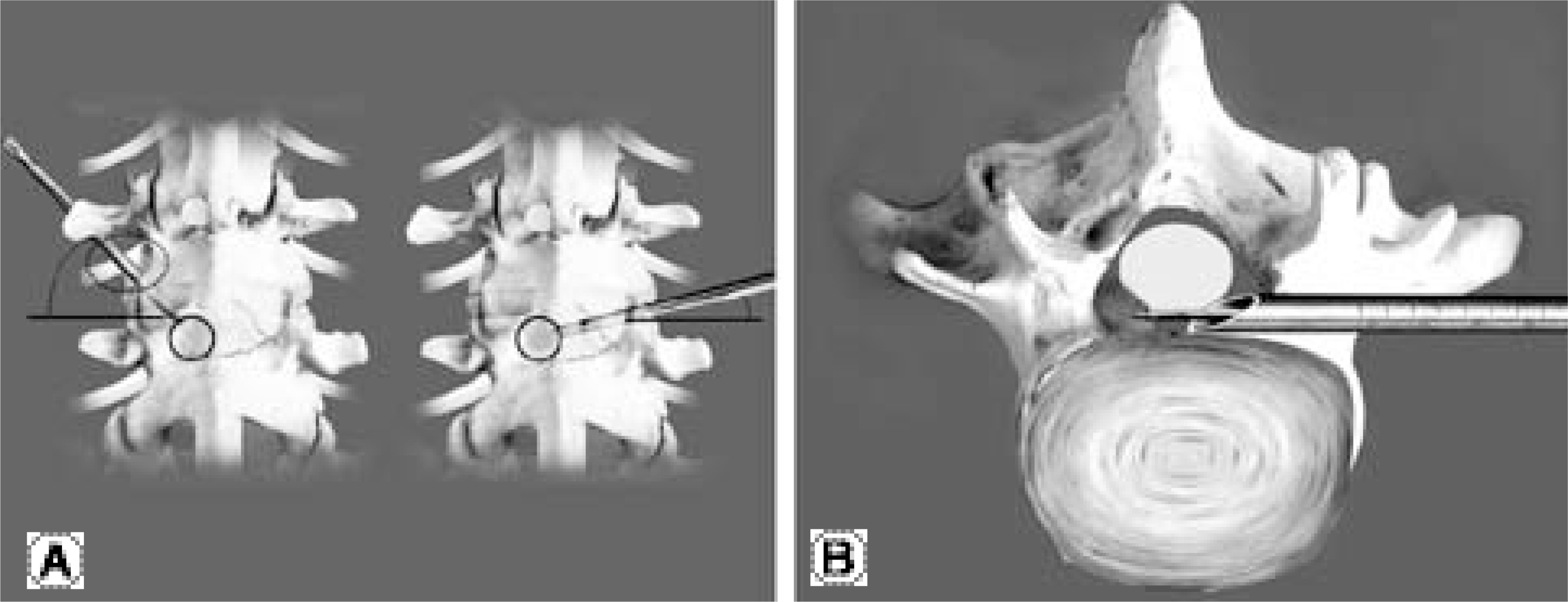
Fig. 10. Diagram of the contralateral transforaminal approach. (A) Contralateral insertion of the obturator and cannula with the low angle than expected. (B) Advance of the cannula not to cross the midline with backward direction of the oblique surface to avoid compression of the central neural structure.
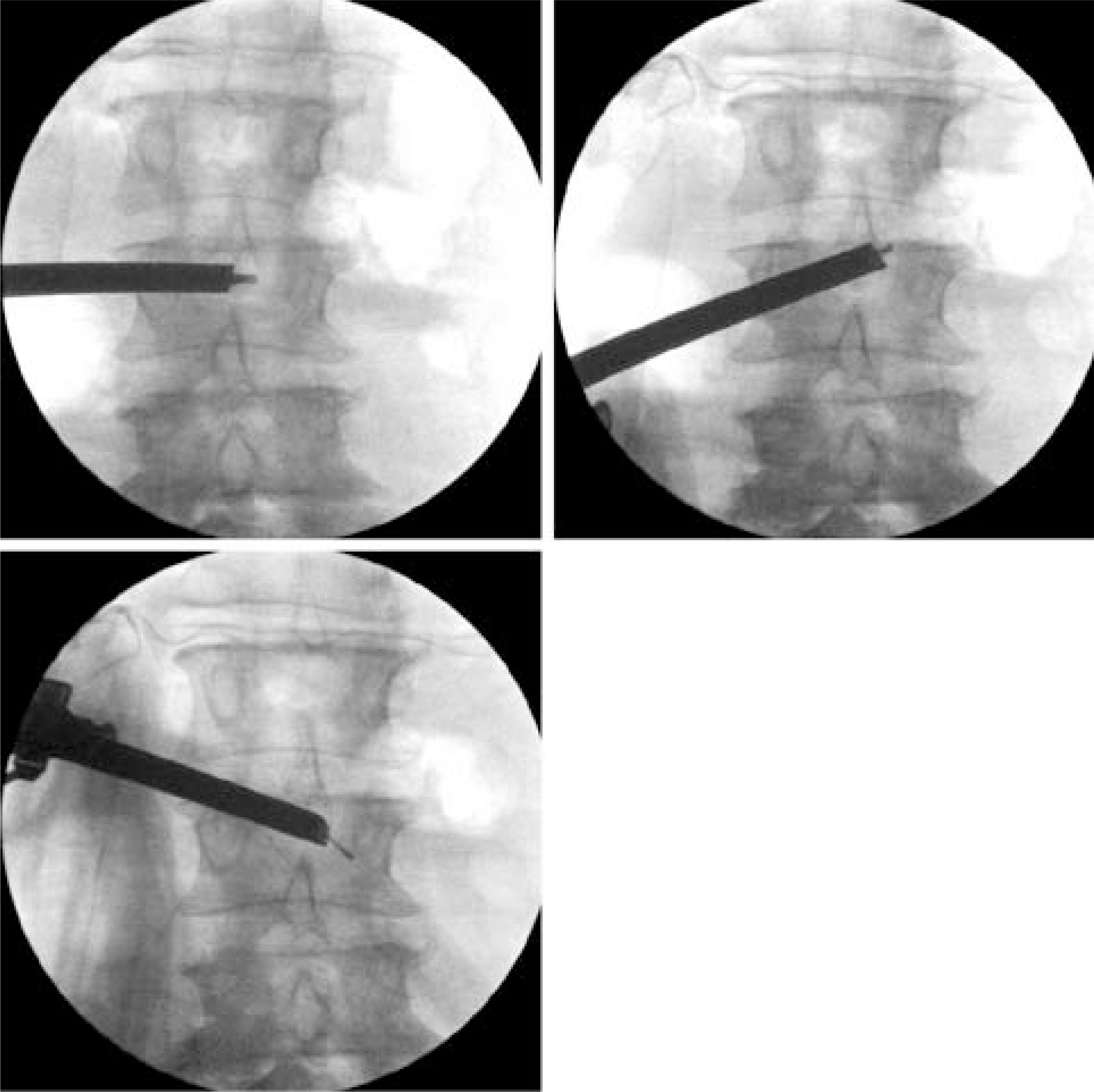
Fig. 11. Fluoroscopic images after insertion of the endoscope through the contralateral interlaminar space.
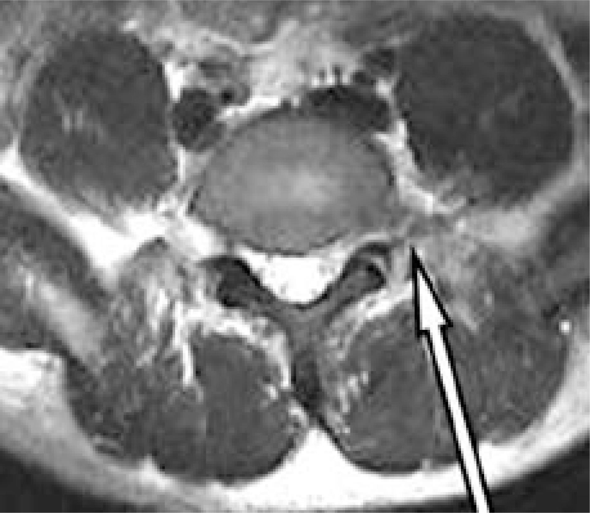
Fig. 12. Intertransverse approaching portal for extraforaminal disc hernation of L5-S1.
Cited by 2 articles
-
Lumbar Discectomy Using Tubular Retractor and Microendoscopy
Sung Chan Ki, Yong Soo Choi, Ki Soo Kim, Woo Jong Kuk
J Korean Soc Spine Surg. 2008;15(4):265-271. doi: 10.7469/JKSS.2008.15.4.265.Comparative Evaluation of Percutaneous Endoscopic Discectomy and Microdiscectomy Using Tubular Retractor System at L4-5 Level
Eui-Chan Jang, Kwang-Sup Song, Ki-Ser Kang, Jae-Yoon Kim, Ki-Seong Kim, Jae June Yang, Young-Bong Ko
J Korean Soc Spine Surg. 2009;16(3):186-193. doi: 10.4184/jkss.2009.16.3.186.
Reference
-
1). Casper W. CA new surgical procedure for lumbar disc herniation causing less tissue damage through a microsurgical approach. Adv Neurosurg. 1977; 4:74–77.2). Williams RW. Microlumbar discectomy: a conservative surgical approach to the virgin herniated lumbar disc. Spine. 1978; 3:175–182.3). Hijikata S, Yamagishi M, Nakayama T. Percutaneous nucleotomy: a new treatment method for lumbar disc herniation. J Toden Hosp. 1975; 5:5–13.4). McCulloch JA, Young PH. Essentials of spinal microsurgery. Philadelphia: Lippincott-Raven;1998.5). Kambin P, Gellmann H. Percutaneous lateral discectomy of the lumbar spine. Clin Orthop. 1983; 174:127–131.
Article6). Stu¨cker R, Krug CH, Reichelt A. Der perkutane trans-foraminale Zugang zum Epiduralraum. Orthopade. 1997; 26:280–287.7). Krugluger J. Transforaminal endoscopic discectomy. (in. Mayer HM, editor. Minimally invasive spine surgery 2nd ed. Springer;p. 315–321. 2005.
Article8). Krugluger J, Knahr K. Minimal invasive disc surgery: a review. Int Orthop. 2001; 24:303–306.9). Rutten S, Mayer O, Godolias G. Endoscopic surgery of the lumbar epidural space (epiduroscopy): results of therapeutic intervention in 93 patients. Minim Invasive Neurosurg. 2003; 2:1018–1022.
Article10). Rutten S. The full-endoscopic interlaminar approach for lumbar disc herniation. (in. Mayer HM, editor. Minimally invasive spine surgery. 2nd ed.Springer;p. 346–355. 2005.
- Full Text Links
-
- Actions
-
Cited
- CITED
-
- Close
- Share
-
- Similar articles
-
- Percutaneous Endoscopic Lumbar Discectomy (PELD)
- Endoscopic Discectomy for the Cauda Equina Syndrome During Third Trimester of Pregnancy
- Clinical Results of Percutaneous Endoscopic Discectomy in Herniated Intervertebral disc of Lumbar Spine
- Far Lateral Extraforaminal Disc Herniation after Percutaneous Laser Lumbar Discectomy
- Revisional Percutaneous Full Endoscopic Disc Surgery for Recurrent Herniation of Previous Open Lumbar Discectomy
