Korean J Obstet Gynecol.
2012 Jun;55(6):413-418. 10.5468/KJOG.2012.55.6.413.
Complete hydatidiform mole with a coexistent fetus diagnosed by magnetic resonance imaging
- Affiliations
-
- 1Department of Obstetrics and Gynecology, Chungbuk National University Hospital, Cheongju, Korea. jeongmed@chungbuk.ac.kr
- 2Medical Research Institute, Chungbuk National University College of Medicine, Cheongju, Korea.
- KMID: 1783935
- DOI: http://doi.org/10.5468/KJOG.2012.55.6.413
Abstract
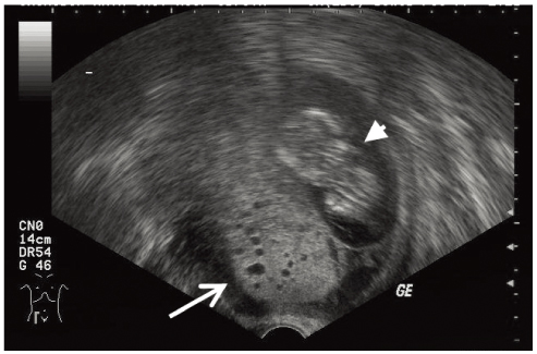
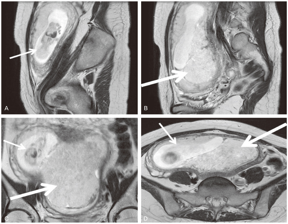
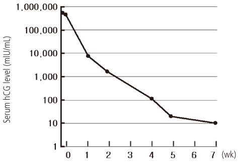
- Hydatidiform mole with coexistent fetus is very rare, arising about 1 in 20,000 to 100,000 pregnancies. There are limited data to guide the management and treatment. Also it is a dilemma to decide continuation or termination of pregnancy. We experienced a case of hydatidiform mole with a coexistent fetus which was diagnosed with magnetic resonance imaging in a woman of 13th weeks of pregnancy. After termination of pregnancy, the patient treated with prophylactic chemotherapy. We report the case with a brief review of literature.
Keyword
MeSH Terms
Figure
Reference
-
1. Steller MA, Genest DR, Bernstein MR, Lage JM, Goldstein DP, Berkowitz RS. Natural history of twin pregnancy with complete hydatidiform mole and coexisting fetus. Obstet Gynecol. 1994. 83:35–42.2. Novick MK, Dillon EH, Epstein NF. AJR teaching file: pregnant woman with vaginal spotting, nausea, and vomiting. AJR Am J Roentgenol. 2010. 194:S79–S82.3. Wee L, Jauniaux E. Prenatal diagnosis and management of twin pregnancies complicated by a co-existing molar pregnancy. Prenat Diagn. 2005. 25:772–776.4. Smith HO, Wiggins C, Verschraegen CF, Cole LW, Greene HM, Muller CY, et al. Changing trends in gestational trophoblastic disease. J Reprod Med. 2006. 51:777–784.5. Kim SJ, Lee C, Kwon SY, Na YJ, Oh YK, Kim CJ. Studying changes in the incidence, diagnosis and management of GTD: the South Korean model. J Reprod Med. 2004. 49:643–654.6. Vejerslev LO. Clinical management and diagnostic possibilities in hydatidiform mole with coexistent fetus. Obstet Gynecol Surv. 1991. 46:577–588.7. Niemann I, Petersen LK, Hansen ES, Sunde L. Predictors of low risk of persistent trophoblastic disease in molar pregnancies. Obstet Gynecol. 2006. 107:1006–1011.8. Sebire NJ, Foskett M, Paradinas FJ, Fisher RA, Francis RJ, Short D, et al. Outcome of twin pregnancies with complete hydatidiform mole and healthy co-twin. Lancet. 2002. 359:2165–2166.9. Garner EI, Goldstein DP, Feltmate CM, Berkowitz RS. Gestational trophoblastic disease. Clin Obstet Gynecol. 2007. 50:112–122.10. Menczer J, Modan M, Serr DM. Prospective follow-up of patients with hydatidiform mole. Obstet Gynecol. 1980. 55:346–349.11. Fowler DJ, Lindsay I, Seckl MJ, Sebire NJ. Routine pre-evacuation ultrasound diagnosis of hydatidiform mole: experience of more than 1000 cases from a regional referral center. Ultrasound Obstet Gynecol. 2006. 27:56–60.12. Shellock FG, Kanal E. SMRI Safety Committee. Policies, guidelines, and recommendations for MR imaging safety and patient management. J Magn Reson Imaging. 1991. 1:97–101.13. Wu TC, Shen SH, Chang SP, Chang CY, Guo WY. Magnetic resonance experience of a twin pregnancy with a normal fetus and hydatidiform mole: a case report. J Comput Assist Tomogr. 2005. 29:415–417.
- Full Text Links
- Actions
-
Cited
- CITED
-
- Close
- Share
- Similar articles
-
- A Case of Complete Hydatidiform Mole in a triplet pregnancy following In Vitro Fertilization and Embryo Transfer
- A Case of Partial Hydatidiform Mole with a Coexistent Live Fetus
- A case of Complete Hydatidiform Mole with a Surviving Coexistent Live Fetus
- A Case of Twin Pregnancy Consisting of Complete Hydatidiform Mole and a Coexisting Fetus Following IUI
- A Case of Complete Hydatidiform Mole with Coexistent Live Fetus


