J Korean Med Sci.
2011 Feb;26(2):237-242. 10.3346/jkms.2011.26.2.237.
Malignancy Rate in Sonographically Suspicious Thyroid Nodules of Less than a Centimeter in Size Does Not Decrease with Decreasing Size
- Affiliations
-
- 1Department of Internal Medicine, Seoul National University College of Medicine, Seoul, Korea. yjparkmd@snu.ac.kr
- 2Department of Internal Medicine, Seoul National University Bundang Hospital, Seongnam, Korea.
- 3Department of Internal Medicine, Seoul National University Hospital Healthcare System, Seoul, Korea.
- KMID: 1782112
- DOI: http://doi.org/10.3346/jkms.2011.26.2.237
Abstract
- We evaluated the malignancy and nondiagnostic rates using fine needle aspiration cytology (FNAC) results in thyroid nodules smaller than 1 cm according to the subdivided size. We retrospectively reviewed the medical records of all subjects underwent FNAC from 2003 to 2009 in our hospital, and 2,756 patients of subcentimeter thyroid nodules with one or more suspicious sonographic features and 7,105 with nodule sized 1 cm or more were included. The malignancy rate was higher in those subcentimeter nodules with suspicious sonographic findings than the nodule sized 1cm or more (19.7% vs 7.8%, P < 0.001). We grouped the nodules based on size with mm interval and observed that the malignancy rate did not decrease but the nondiagnostic results increased its size decrement. When we divided the subjects arbitrarily into a 5 mm or smaller and a 6-9 mm sized group, nondiagnostic cytology findings were reported more frequently in the smaller group (24.3% vs 18.1%, P = 0.001), while the rate of "malignant" was similar (18.3% vs 15.5%, P = 0.123) and the rate of "suspicious for malignancy" was higher (6.8% vs 2.9%, P < 0.001). Therefore when we decide to perform FNAC or not in subcentimeter-sized nodules, we should consider sonographic findings and other clinical risk factors but not the nodular size itself.
MeSH Terms
Figure
-
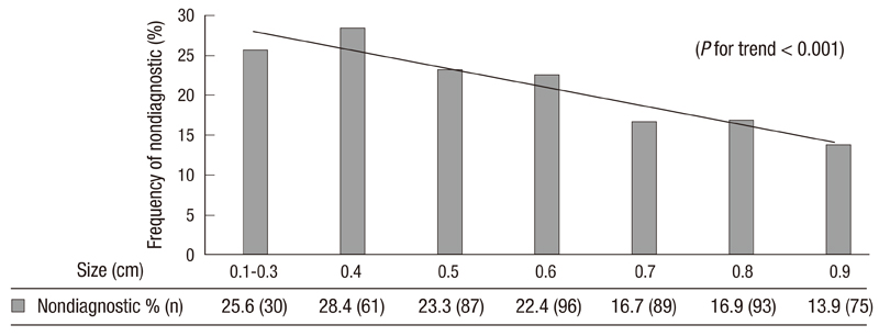
Fig. 1 The nondiagnostic rate of FNAC was increased by decreasing nodules in subcentimeter sized thyroid nodules.
Reference
-
1. Ezzat S, Sarti DA, Cain DR, Braunstein GD. Thyroid incidentalomas. Prevalence by palpation and ultrasonography. Arch Intern Med. 1994. 154:1838–1840.2. Jung KW, Won YJ, Park S, Kong HJ, Sung J, Shin HR, Park EC, Lee JS. Cancer statistics in Korea: incidence, mortality and survival in 2005. J Korean Med Sci. 2009. 24:995–1003.3. Jung KW, Park S, Kong HJ, Won YJ, Boo YK, Shin HR, Park EC, Lee JS. Cancer statistics in Korea: incidence, mortality and survival in 2006-2007. J Korean Med Sci. 2010. 25:1113–1121.4. Wada N, Duh QY, Sugino K, Iwasaki H, Kameyama K, Mimura T, Ito K, Takami H, Takanashi Y. Lymph node metastasis from 259 papillary thyroid microcarcinomas: frequency, pattern of occurrence and recurrence, and optimal strategy for neck dissection. Ann Surg. 2003. 237:399–407.5. Lee NS, Bae JS, Jeong SR, Jung CK, Lim DJ, Park WC, Kim JS, Kim SN. Risk factors of lymph node metastasis in papillary thyroid microcarcinoma. J Korean Surg Soc. 2010. 78:82–86.6. Noguchi S, Yamashita H, Uchino S, Watanabe S. Papillary microcarcinoma. World J Surg. 2008. 32:747–753.7. Kim EK, Park CS, Chung WY, Oh KK, Kim DI, Lee JT, Yoo HS. New sonographic criteria for recommending fine-needle aspiration biopsy of nonpalpable solid nodules of the thyroid. AJR Am J Roentgenol. 2002. 178:687–691.8. Cibas ES, Ali SZ. The Bethesda System for Reporting Thyroid Cytopathology. Thyroid. 2009. 19:1159–1165.9. Shimura H, Haraguchi K, Hiejima Y, Fukunari N, Fujimoto Y, Katagiri M, Koyanagi N, Kurita T, Miyakawa M, Miyamoto Y, Suzuki N, Suzuki S, Kanbe M, Kato Y, Murakami T, Tohno E, Tsunoda-Shimizu H, Yamada K, Ueno E, Kobayashi K, Kobayashi T, Yokozawa T, Kitaoka M. Distinct diagnostic criteria for ultrasonographic examination of papillary thyroid carcinoma: a multicenter study. Thyroid. 2005. 15:251–258.10. Simpson WJ, McKinney SE, Carruthers JS, Gospodarowicz MK, Sutcliffe SB, Panzarella T. Papillary and follicular thyroid cancer. Prognostic factors in 1,578 patients. Am J Med. 1987. 83:479–488.11. Bilimoria KY, Bentrem DJ, Ko CY, Stewart AK, Winchester DP, Talamonti MS, Sturgeon C. Extent of surgery affects survival for papillary thyroid cancer. Ann Surg. 2007. 246:375–381.12. Roti E, degli Uberti EC, Bondanelli M, Braverman LE. Thyroid papillary microcarcinoma: a descriptive and meta-analysis study. Eur J Endocrinol. 2008. 159:659–673.13. Ito Y, Uruno T, Nakano K, Takamura Y, Miya A, Kobayashi K, Yokozawa T, Matsuzuka F, Kuma S, Kuma K, Miyauchi A. An observation trial without surgical treatment in patients with papillary microcarcinoma of the thyroid. Thyroid. 2003. 13:381–387.14. Park YJ, Kim YA, Lee YJ, Kim SH, Park SY, Kim KW, Chung JK, Youn YK, Kim KH, Park do J, Cho BY. Papillary microcarcinoma in comparison with larger papillary thyroid carcinoma in BRAF(V600E) mutation, clinicopathological features, and immunohistochemical findings. Head Neck. 2010. 32:38–45.15. Park YJ, Kwak SH, Kim DC, Kim H, Choe G, Park do J, Jang HC, Park SH, Cho BY, Park SY. Diagnostic value of galectin-3, HBME-1, cytokeratin 19, high molecular weight cytokeratin, cyclin D1 and p27(kip1) in the differential diagnosis of thyroid nodules. J Korean Med Sci. 2007. 22:621–628.16. Chow SM, Law SC, Chan JK, Au SK, Yau S, Lau WH. Papillary microcarcinoma of the thyroid-Prognostic significance of lymph node metastasis and multifocality. Cancer. 2003. 98:31–40.17. Roti E, Rossi R, Trasforini G, Bertelli F, Ambrosio MR, Busutti L, Pearce EN, Braverman LE, Degli Uberti EC. Clinical and histological characteristics of papillary thyroid microcarcinoma: results of a retrospective study in 243 patients. J Clin Endocrinol Metab. 2006. 91:2171–2178.18. Berker D, Aydin Y, Ustun I, Gul K, Tutuncu Y, Işik S, Delibasi T, Guler S. The value of fine-needle aspiration biopsy in subcentimeter thyroid nodules. Thyroid. 2008. 18:603–608.19. Kim SJ, Kim EK, Park CS, Chung WY, Oh KK, Yoo HS. Ultrasound-guided fine-needle aspiration biopsy in nonpalpable thyroid nodules: is it useful in infracentimetric nodules? Yonsei Med J. 2003. 44:635–640.20. Kim DW, Lee EJ, Kim SH, Kim TH, Lee SH, Kim DH, Rho MH. Ultrasound-guided fine-needle aspiration biopsy of thyroid nodules: comparison in efficacy according to nodule size. Thyroid. 2009. 19:27–31.21. Nam-Goong IS, Kim HY, Gong G, Lee HK, Hong SJ, Kim WB, Shong YK. Ultrasonography-guided fine-needle aspiration of thyroid incidentaloma: correlation with pathological findings. Clin Endocrinol (Oxf). 2004. 60:21–28.22. Rosen IB, Azadian A, Walfish PG, Salem S, Lansdown E, Bedard YC. Ultrasound-guided fine-needle aspiration biopsy in the management of thyroid disease. Am J Surg. 1993. 166:346–349.23. Redman R, Zalaznick H, Mazzaferri EL, Massoll NA. The impact of assessing specimen adequacy and number of needle passes for fine-needle aspiration biopsy of thyroid nodules. Thyroid. 2006. 16:55–60.24. Leenhardt L, Hejblum G, Franc B, Fediaevsky LD, Delbot T, Le Guillouzic D, Ménégaux F, Guillausseau C, Hoang C, Turpin G, Aurengo A. Indications and limits of ultrasound-guided cytology in the management of nonpalpable thyroid nodules. J Clin Endocrinol Metab. 1999. 84:24–28.
- Full Text Links
-
- Actions
-
Cited
- CITED
-
- Close
- Share
-
- Similar articles
-
- Updated guidelines for the diagnosis and management of thyroid nodules
- Mixed Echoic Thyroid Nodules on Ultrasound: Approach to Management
- Subcategorization of intermediate suspicion thyroid nodules based on suspicious ultrasonographic findings
- Risk of Malignancy in Thyroid Nodules 4 cm or Larger
- Impact of Nodule Size on Malignancy Risk Differs according to the Ultrasonography Pattern of Thyroid Nodules
