What are the Causative Factors for a Slow, Progressive Enlargement of a Chronic Subdural Hematoma?
- Affiliations
-
- 1Department of Neurosurgery, Inha University College of Medicine, Incheon, Korea. nspco@inha.ac.kr
- KMID: 1779522
- DOI: http://doi.org/10.3349/ymj.2007.48.2.210
Abstract
- PURPOSE
To test the hypothesis that chronic subdural hematoma (CSDH) enlarges by the causative factors, this study has performed. METERIALS AND METHODS: In 10 patients with CSDH, coagulation factors in venous blood taken at the time of surgery and hematomic contents aspirated from the CSDH were studied, using both laboratory assays and microscopy. RESULTS: When compared to the range of normal plasma, the hematoma fluids demonstrated a marked reduction in factor II, V, VII, VIII, and X, moderate reduction of factors IX and XI, and slight reduction of factor XII. Activated protein C and antithrombin III levels were decreased. The FDP (Fibrinogen Degradation Product) levels in chronic subdural hematoma were extremely high. The endothelial cells of the macrocapillaries (also called "sinusoid") showed numerous gap junctions between adjacent endothelial cells and a thinness or absence of the basement membrane, suggesting that the macrocapillaries are very fragile and susceptible to bleeding. CONCLUSION: Excessive coagulation in the hematoma, predominantly via the extrinsic clotting pathway, local hyperfibrinolysis, transmitted pulsations, and characteristics of the macrocapillaries play an important role in the leakage of blood and the enlargement of CSDH.
Keyword
Figure
-
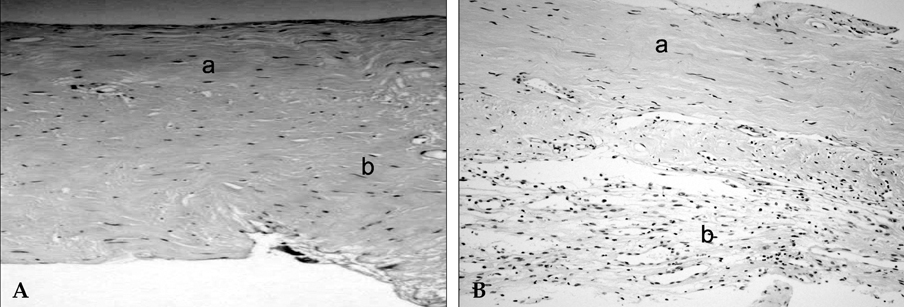
Fig. 1 Photomicrographs of the dura mater. (A) The Compact lamella contains the tight fibrous tissue with fibroblasts, collagen of the meningeal layer(a), the loose lamellas contains the loose fibrous tissue with a few blood vessels(b). (B) Photographs of the outer membrane of subdural caopsule. Note the connective tissue proliferation at the outer side of the outer membrane (compact fibrous tissue, a) and multiple neo-vascular sinusoidal channels overlying fresh red blood cells (loose fibrous tissue; vascular layer, b) ×100.

Fig. 2 Electron micrograph of the external membrane of a subdural hematma showing a macrocapillary. Its lumen is as large as 40m in diameter with erythrocytes (Ec) and consisted of single layer of flattened endothelial cells with a faint or discontinuous basement membrane. Endothelial cells have hyper-chromatic, large nucleus with a high nucleus/cytoplasm ratio. There are two erythrocytes at the outside of the capillary wall. P, pseudopod-like protrusion; N, nucleus; L, lumen (×5,000).

Fig. 3 Electron micrograph of a portion of a macrocapillary with a endothelial gap junction. The endothelal gap junction is as large as 0.6m in diameter. Basement membrane is discontinuous and faint. Two extravasated erythrocytes can be seen. The endothelial cytoplasm contains many ribosomes. L, lumen; Ec, erythrocyte (×10,000).
Cited by 1 articles
-
The Relationship between Postoperative Drainage Volume and Brain Shifting Index in Chronic Subdural Hematoma
Chang-Hyun Oh, Dong-Keun Hyun, Seung-Hwan Yoon, Hyeon-Seon Park, Eunyoung Kim, Hyeong-Chun Park, Chong-Oon Park
J Korean Neurotraumatol Soc. 2008;4(2):70-76. doi: 10.13004/jknts.2008.4.2.70.
Reference
-
1. Markwalder TM. Chronic subdural hematomas: a review. J Neurosurg. 1981. 54:637–645.2. Friede RL, Schachenmayr W. The origin of subdural neomembranes. II. Fine structural of neomembranes. Am J Pathol. 1978. 92:69–84.3. Sato S, Suzuki J. Ultrastructural observations of the capsule of chronic subdural hematoma in various clinical stages. J Neurosurg. 1975. 43:569–578.4. Yamashima T, Yamamoto S. How do vessels proliferate in the capsule of a chronic subdural hematoma? Neurosurgery. 1984. 15:672–678.5. Ito H, Yamamoto S, Komai T, mizukoshi H. Role of local hyperfibrinolysis in the etiology of chronic subdural hematoma. J Neurosurg. 1976. 45:26–31.6. Ito H, Komai T, Yamamoto S. Fibrinolytic enzyme in the lining walls of chronic subdural hematoma. J Neurosurg. 1978. 48:197–200.7. Weir B, Gordon P. Factors affecting coagulation. Fibrinolysis in chronic subdural fluid collections. J Neurosurg. 1983. 58:242–245.8. Dati F, Barthels M, Conard J, Fluckiger J, Girolami A, Hanseler E, et al. Multicenter evaluation of a chromogenic substrate method for photometric determination of prothrombin time. Thromb Haemost. 1987. 58:856–865.9. Kawakami Y, Tanimoto T, Shimamura Y. Coagulopathy in chronic subdural hematoma. Neurol Med Chir (Tokyo). 1991. 31:32–36.10. Martinoli JL, Stocker K. Fast functional protein C assay using Protac, a novel protein C activator. Thromb Res. 1986. 43:253–264.11. Scully MF, Kakkar VV. Methods for semi micro or automated determination of thrombin, antithrombin, and heparin cofactor using he substrate, H-D-Phe-Pip-Arg-p-nitroanilide-2HCL. Clin Chim Acta. 1977. 79:595–602.12. Hauw J, Berger B, Escourolle R. Electron Microscopic study of the developing capillaries of human brain. Acta Neuropathol(Berl). 1975. 31:229–242.13. Putamen J, Cushing H. Chronic subdural hematoma. Its pathology, its relation to pachymeningitis hemorrhagica and its surgical treatment. Arch Surg. 1925. 11:329–393.14. Rhodin JA. Ultrastructure of mammalian venous capillaries, venules, and small collecting veins. J Ultrastruct Res. 1968. 25:452–500.15. Yamashima T, Yamamoto S, Friede RL. The role of endothelial gap junctions in the enlargement of chronic subdural hematomas. J Neurosurg. 1983. 59:298–303.16. Yamamoto T, Sato S. Electron microscope studies on the vessel wall and the leukocyte emigration. Arch Histol Jpn. 1966. 27:297–310.17. Scotti G, Terbrugge K, Melancon D, Belanger G. Evaluation of the age of subdural hematomas by computerized tomography. J Neurosurg. 1977. 47:311–315.18. Rowley DA. Venous constriction as the cause of increased vascular permeability produced by 5-hydroxytryptamine, histamine, bradykinin, and 48/80 in the rat. Br J Exp Pathol. 1964. 45:56–67.19. Watanabe S, Shimada H, Ishii S. Production of clinical form of chronic subdural hematoma in experimental animals. J Neurosurg. 1972. 37:552–561.20. Yamashima T, Shimoji T, Komai T, Kubota T, Ito H, Yamamoto S. Growing mechanism of chronic subdural hematoma-light and electron microscopic study on outer membranes of chronic subdural hematoma (author's transl). Neurol Med Chir(Tokyo). 1978. 18:743–752.21. Handin RI. Braunwald E, Fauci A, Kasper DL, Hauser S, Longo DL, Jameson JL, editors. Bleeding and thrombosis. Harrison's Principles of Internal Medicine. 2005. 16th ed. New York: McGraw-Hill;337–343.22. Murakami H, Hirose Y, Sagoh M, Shimizu K, Kojima M, Gotoh K, et al. Why do chronic subdural hematomas continue to grow slowly any coagulate? Role of thrombomodulin in the mechanism. J Neurosurg. 2002. 96:877–884.
- Full Text Links
-
- Actions
-
Cited
- CITED
-
- Close
- Share
-
- Similar articles
-
- Chronic Subdural Hematoma Superimposed on Posttraumatic Subdural Hygroma: A Report of Three Cases
- Bilateral Acute Subdural Hematoma Following Evacuation of Chronic Subdural Hematoma
- Arachnoid Cyst with Spontaneous Intracystic Hemorrhage and Chronic Subdural Hematoma
- Treatment of Chronic Subdural Hematoma with Arachnoid Cyst
- Prognostic Factors of Chronic Subdural Hematoma
