J Korean Med Sci.
2013 May;28(5):709-716. 10.3346/jkms.2013.28.5.709.
Association of Apolipoprotein B/Apolipoprotein A1 Ratio and Coronary Artery Stenosis and Plaques Detected by Multi-Detector Computed Tomography in Healthy Population
- Affiliations
-
- 1Department of Internal Medicine, Asan Medical Center, University of Ulsan College of Medicine, Seoul, Korea. lwjatlas@naver.com
- 2Health Screening and Promotion Center, Asan Medical Center, University of Ulsan College of Medicine, Seoul, Korea.
- 3Department of Radiology, Asan Medical Center, University of Ulsan College of Medicine, Seoul, Korea.
- KMID: 1777558
- DOI: http://doi.org/10.3346/jkms.2013.28.5.709
Abstract
- Despite the noninvasiveness and accuracy of multidetector computed tomography (MDCT), its use as a routine screening tool for occult coronary atherosclerosis is unclear. We investigated whether the ratio of apolipoprotein B (apoB) to apolipoprotein A1 (apoA1), an indicator of the balance between atherogenic and atheroprotective cholesterol transport could predict occult coronary atherosclerosis detected by MDCT. We collected the data of 1,401 subjects (877 men and 524 women) who participated in a routine health screening examination of Asan Medical Center. Significant coronary artery stenosis defined as > 50% stenosis was detected in 114 subjects (8.1%). An increase in apoB/A1 quartiles was associated with increased percentages of subjects with significant coronary stenosis and noncalcified plaques (NCAP). After adjustment for confounding variables, each 0.1 increase in serum apoB/A1 was significantly associated with increased odds ratios (ORs) for coronary stenosis and NCAP of 1.23 and 1.18, respectively. The optimal apoB/A1 ratio cut off value for MDCT detection of significant coronary stenosis was 0.58, which had a sensitivity of 70.2% and a specificity of 48.2% (area under the curve, 0.61; 95% CI, 0.58-0.63, P < 0.001). Our results indicate that apoB/A1 ratio is a good indicator of occult coronary atherosclerosis detected by coronary MDCT.
MeSH Terms
Figure
-
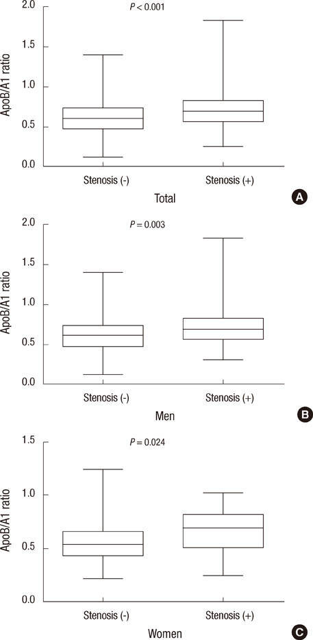
Fig. 1 Box-whisker plots of apoB/A1 ratios according to the presence of significant coronary stenosis in total subjects (A), men (B), and women (C). The ends of each whisker indicate the 5th and 95th percentiles of apoB/A1 ratio.
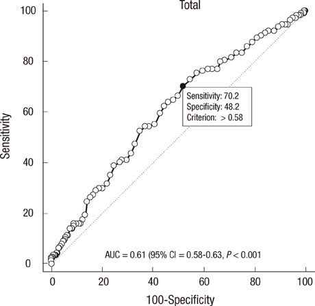
Fig. 2 Receiver operating characteristic (ROC) curve and optimal apoB/A1 ratio cutoff value (•) for detecting significant coronary stenosis by multidetector computed tomography (MDCT).
Reference
-
1. Expert Panel on Detection, Evaluation, and Treatment of High Blood Cholesterol in Adults. Executive Summary of the Third Report of the National Cholesterol Education Program (NCEP) Expert Panel on Detection, Evaluation, and Treatment of High Blood Cholesterol in Adults (Adult Treatment Panel III). JAMA. 2001. 285:2486–2497.2. Marcovina S, Packard CJ. Measurement and meaning of apolipoprotein AI and apolipoprotein B plasma levels. J Intern Med. 2006. 259:437–446.3. Yusuf S, Hawken S, Ounpuu S, Dans T, Avezum A, Lanas F, McQueen M, Budaj A, Pais P, Varigos J, et al. Effect of potentially modifiable risk factors associated with myocardial infarction in 52 countries (the INTERHEART Study): case-control study. Lancet. 2004. 364:937–952.4. Budoff MJ, Achenbach S, Blumenthal RS, Carr JJ, Goldin JG, Greenland P, Guerci AD, Lima JA, Rader DJ, Rubin GD, et al. Assessment of coronary artery disease by cardiac computed tomography: a scientific statement from the American Heart Association Committee on Cardiovascular Imaging and Intervention, Council on Cardiovascular Radiology and Intervention, and Committee on Cardiac Imaging, Council on Clinical Cardiology. Circulation. 2006. 114:1761–1791.5. Foster G, Shah H, Sarraf G, Ahmadi N, Budoff M. Detection of noncalcified and mixed plaque by multirow detector computed tomography. Expert Rev Cardiovasc Ther. 2009. 7:57–64.6. Achenbach S, Daniel WG. Current role of cardiac computed tomography. Herz. 2007. 32:97–107.7. Peppa M, Betsi G, Dimitriadis G. Lipid abnormalities and cardiometabolic risk in patients with overt and subclinical thyroid disease. J Lipids. 2011. 2011:575840.8. Seidel D. Lipoproteins in liver disease. J Clin Chem Clin Biochem. 1987. 25:541–551.9. Batista MC, Welty FK, Diffenderfer MR, Sarnak MJ, Schaefer EJ, Lamon-Fava S, Asztalos BF, Dolnikowski GG, Brousseau ME, Marsh JB. Apolipoprotein A-I, B-100, and B-48 metabolism in subjects with chronic kidney disease, obesity, and the metabolic syndrome. Metabolism. 2004. 53:1255–1261.10. Austen WG, Edwards JE, Frye RL, Gensini GG, Gott VL, Griffith LS, McGoon DC, Murphy ML, Roe BB. A reporting system on patients evaluated for coronary artery disease: report of the Ad Hoc Committee for Grading of Coronary Artery Disease, Council on Cardiovascular Surgery, American Heart Association. Circulation. 1975. 51:5–40.11. Leber AW, Becker A, Knez A, von Ziegler F, Sirol M, Nikolaou K, Ohnesorge B, Fayad ZA, Becker CR, Reiser M, et al. Accuracy of 64-slice computed tomography to classify and quantify plaque volumes in the proximal coronary system: a comparative study using intravascular ultrasound. J Am Coll Cardiol. 2006. 47:672–677.12. Agatston AS, Janowitz WR, Hildner FJ, Zusmer NR, Viamonte M Jr, Detrano R. Quantification of coronary artery calcium using ultrafast computed tomography. J Am Coll Cardiol. 1990. 15:827–832.13. Badimon JJ, Ibanez B, Cimmino G. Genesis and dynamics of atherosclerotic lesions: implications for early detection. Cerebrovasc Dis. 2009. 27:38–47.14. Holmes DR Jr, Elveback LR, Frye RL, Kottke BA, Ellefson RD. Association of risk factor variables and coronary artery disease documented with angiography. Circulation. 1981. 63:293–299.15. Walldius G, Jungner I, Holme I, Aastveit AH, Kolar W, Steiner E. High apolipoprotein B, low apolipoprotein A-I, and improvement in the prediction of fatal myocardial infarction (AMORIS study): a prospective study. Lancet. 2001. 358:2026–2033.16. Noma A, Yokosuka T, Kitamura K. Plasma lipids and apolipoproteins as discriminators for presence and severity of angiographically defined coronary artery disease. Atherosclerosis. 1983. 49:1–7.17. Reinhart RA, Gani K, Arndt MR, Broste SK. Apolipoproteins A-I and B as predictors of angiographically defined coronary artery disease. Arch Intern Med. 1990. 150:1629–1633.18. Enkhmaa B, Anuurad E, Zhang Z, Pearson TA, Berglund L. Usefulness of apolipoprotein B/apolipoprotein A-I ratio to predict coronary artery disease independent of the metabolic syndrome in African Americans. Am J Cardiol. 2010. 106:1264–1269.19. Thaulow E, Erikssen J, Sandvik L, Erikssen G, Jorgensen L, Cohn PF. Initial clinical presentation of cardiac disease in asymptomatic men with silent myocardial ischemia and angiographically documented coronary artery disease (the Oslo Ischemia Study). Am J Cardiol. 1993. 72:629–633.20. Pilote L, Pashkow F, Thomas JD, Snader CE, Harvey SA, Marwick TH, Lauer MS. Clinical yield and cost of exercise treadmill testing to screen for coronary artery disease in asymptomatic adults. Am J Cardiol. 1998. 81:219–224.21. Choi EK, Choi SI, Rivera JJ, Nasir K, Chang SA, Chun EJ, Kim HK, Choi DJ, Blumenthal RS, Chang HJ. Coronary computed tomography angiography as a screening tool for the detection of occult coronary artery disease in asymptomatic individuals. J Am Coll Cardiol. 2008. 52:357–365.22. Schuijf JD, van Werkhoven JM, Pundziute G, Jukema JW, Decramer I, Stokkel MP, Dibbets-Schneider P, Schalij MJ, Reiber JH, van der Wall EE, et al. Invasive versus noninvasive evaluation of coronary artery disease. JACC Cardiovasc Imaging. 2008. 1:190–199.23. Vanhoenacker PK, Heijenbrok-Kal MH, Van Heste R, Decramer I, Van Hoe LR, Wijns W, Hunink MG. Diagnostic performance of multidetector CT angiography for assessment of coronary artery disease: meta-analysis. Radiology. 2007. 244:419–428.24. Hacker M, Jakobs T, Hack N, Nikolaou K, Becker C, von Ziegler F, Knez A, König A, Klauss V, Reiser M, et al. Sixty-four slice spiral CT angiography does not predict the functional relevance of coronary artery stenoses in patients with stable angina. Eur J Nucl Med Mol Imaging. 2007. 34:4–10.25. Schuijf JD, Wijns W, Jukema JW, Atsma DE, de Roos A, Lamb HJ, Stokkel MP, Dibbets-Schneider P, Decramer I, De Bondt P, et al. Relationship between noninvasive coronary angiography with multi-slice computed tomography and myocardial perfusion imaging. J Am Coll Cardiol. 2006. 48:2508–2514.26. Nam HJ, Jung IH, Kim J, Kim JH, Suh J, Kim HS, Kim HK, Jung YJ, Kang JW, Lee S. Association between brachial-ankle pulse wave velocity and occult coronary artery disease detected by multi-detector computed tomography. Int J Cardiol. 2012. 157:227–232.27. Rumberger JA, Simons DB, Fitzpatrick LA, Sheedy PF, Schwartz RS. Coronary artery calcium area by electron-beam computed tomography and coronary atherosclerotic plaque area: a histopathologic correlative study. Circulation. 1995. 92:2157–2162.28. Stary HC, Chandler AB, Dinsmore RE, Fuster V, Glagov S, Insull W Jr, Rosenfeld ME, Schwartz CJ, Wagner WD, Wissler RW. A definition of advanced types of atherosclerotic lesions and a histological classification of atherosclerosis: a report from the Committee on Vascular Lesions of the Council on Arteriosclerosis, American Heart Association. Circulation. 1995. 92:1355–1374.29. Hoffmann U, Moselewski F, Nieman K, Jang IK, Ferencik M, Rahman AM, Cury RC, Abbara S, Joneidi-Jafari H, Achenbach S, et al. Noninvasive assessment of plaque morphology and composition in culprit and stable lesions in acute coronary syndrome and stable lesions in stable angina by multidetector computed tomography. J Am Coll Cardiol. 2006. 47:1655–1662.30. Fujii K, Kobayashi Y, Mintz GS, Takebayashi H, Dangas G, Moussa I, Mehran R, Lansky AJ, Kreps E, Collins M, et al. Intravascular ultrasound assessment of ulcerated ruptured plaques: a comparison of culprit and nonculprit lesions of patients with acute coronary syndromes and lesions in patients without acute coronary syndromes. Circulation. 2003. 108:2473–2478.31. Kinlay S, Dobson AJ, Heller RF, Dickeson JE, Ryan S. Lipid and apolipoprotein levels in an Australian community. Med J Aust. 1991. 154:170–175.32. Marcovina SM, Albers JJ, Kennedy H, Mei JV, Henderson LO, Hannon WH. International Federation of Clinical Chemistry standardization project for measurements of apolipoproteins A-I and B: IV. comparability of apolipoprotein B values by use of International Reference Material. Clin Chem. 1994. 40:586–592.33. Waxman S, Ishibashi F, Muller JE. Detection and treatment of vulnerable plaques and vulnerable patients: novel approaches to prevention of coronary events. Circulation. 2006. 114:2390–2411.
- Full Text Links
-
- Actions
-
Cited
- CITED
-
- Close
- Share
-
- Similar articles
-
- Coronary Artery Calcification and Serum Apolipoprotein A-1 in Patients with Type 2 Diabetes
- The Relationship Between Coronary Artery Calcification and Serum Apolipoprotein A-1 in Patients with Type 2 Diabetes
- Three cases of right coronary anomaly confirmed by multi-detector computed tomography
- A Study of Plasma Apolipoprotein A-1 and Apolipoprotein B Levels in Patients with Coronary Artery Disease
- Apolipoprotein B/A1 Ratio as Risk Factor for Cerebral Ischemic Stroke
